
Precise robot radiotherapy system under guidance of MRI

A robotic and precise technology, applied in the field of medical devices, can solve the problems of inability to distinguish the boundary between tumor and normal tissue, poor resolution of soft tissue by X-ray imaging, slow rotation speed, etc., to avoid additional damage and strengthen treatment. ability, effect of small radiation doses

Inactive Publication Date: 2016-01-20
BEIJING JIANLIAN MEDICAL TECH CO LTD
View PDF4 Cites 9 Cited by
  • Summary
  • Abstract
  • Description
  • Claims
  • Application Information

AI Technical Summary

Problems solved by technology

[0003] To ensure that the X / γ-ray beam is irradiated on the target area, certain imaging equipment is required for real-time guidance. Most of the existing image guidance methods in radiotherapy use cone beam CT (CBCT) or planar X-ray imaging equipment for guidance. Here It is collectively referred to as X-ray imaging-guided radiotherapy, and its disadvantages are: (1) From the beginning of diagnosis to the end of treatment, the patient receives X-ray scans of X-ray imager multiple times, and not only the lesion area, but also the non-lesion area will receive additional radiation therapy. The cumulative dose of ionizing radiation has far exceeded the annual allowable dose for normal people, which can cause cancer or new disease-causing problems in patients, especially women and children; (2) The current X-ray imaging-guided radiotherapy , is not real-time, it is difficult to track the treatment situation in three-dimensional space in real time, and it is necessary to use other auxiliary means to establish three-dimensional space coordinates to track the treatment situation, such as the combination of invasive methods of gold particle implantation and planar X-ray imaging. Realize the tracking of the lesion (such as Cyberknife technology), and most of the radiation therapy guided by cone beam CT fixes the cone beam CT system on the X-ray source rotating bracket. Since the rotation speed of the frame is very slow, the rotation angle is only limited In a very small range, it is impossible to form a complete angular reconstruction of the CT data set for the lesion and the target area during the implementation of radiotherapy, so that radiotherapy cannot be guided in real time in three-dimensional space; (3) X-ray imaging has poor resolution of soft tissues, The boundary between tumor and normal tissue cannot be distinguished well; (4) The contrast of X-ray imaging to soft tissue is single, and the effect of treatment can only be judged according to the change of tumor size, but the change of tumor size is often not visible after a course of treatment ; Off-line magnetic resonance imaging equipment has multiple contrast differences such as molecular imaging, and uses special supporting software and methods to realize early judgment of curative effect

Method used


Image

  • Precise robot radiotherapy system under guidance of MRI
  • Precise robot radiotherapy system under guidance of MRI
  • Precise robot radiotherapy system under guidance of MRI

Examples


Embodiment Construction

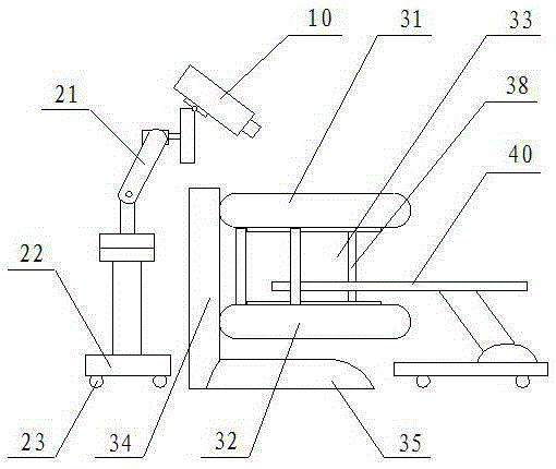
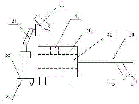
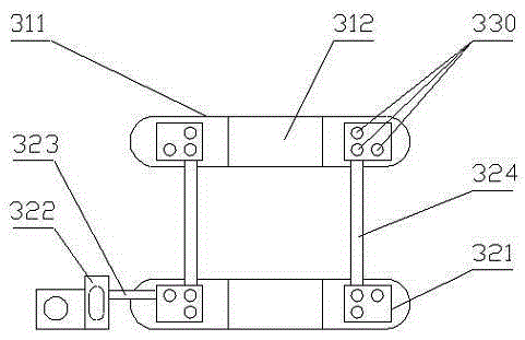
[0010] See figure 1 -3, the present invention provides a MRI-guided robot precise radiotherapy system , Including X / γ-ray sources (including their collimator system) 10, and also an online magnetic resonance imaging device provided with a magnet for forming a magnetic field, and the magnet is preferably a superconducting magnet, The superconducting magnet may be a split superconducting magnet or a cylindrical superconducting magnet 40. The split superconducting magnet is mainly composed of two magnetic poles 31 and 32 corresponding to each other. A gap (spacing) 33 is left, the cylindrical superconducting magnet includes superconducting coils and is provided with an axial hole 42, and the X / γ-ray source is installed on the robot arm, which is driven by the robot Movement (change of position and / or direction). For on-line magnetic resonance imaging equipment using split superconducting magnets, the radiotherapy beam (X-ray beam or γ-ray beam, etc.) emitted by the X / γ-ray source...


PUM


Abstract

The invention relates to a precise robot radiotherapy system under guidance of MRI. The system comprises an x / gamma-ray source and on-line magnetic resonance imaging equipment. The on-line magnetic resonance imaging equipment is provided with a split type / cylindrical superconducting magnet for generating a magnetic field. The x / gamma-ray source is installed on a mechanical arm of a robot and is driven by the robot to move, so that a radiotherapy beam emitted by the x / gamma-ray source can enter a focus imaging region between two magnetic poles of the split type superconducting magnet or inside the cylindrical superconducting magnet through the gap between the magnetic poles and / or holes in the magnetic poles. According to the invention, under guidance of the open type magnetic resonance imaging equipment, a mechanical arm or mechanical arms of one or more robots is / are used for operating one or a plurality of treatment heads to irradiate x / gamma rays on tumor focuses, so that the x / gamma ray therapeutic machine can have more treatment functions for patients, a fast treatment speed, a better treatment capacity, and a good treatment effect.

Description

technical field [0001] The invention relates to an MRI (Magnetic Resonance Imaging)-guided robotic precision radiotherapy system, in particular using an electron linear accelerator / 60 A radiotherapy system for Coγ-rays belongs to the technical field of medical equipment. Background technique [0002] The electron linear accelerator uses electrons with a certain energy to interact with the microwave electric field to obtain higher energy and form an electron beam with a certain energy. The electron beam is directly drawn from the electron gun to bombard the target area such as the tumor, which is called electron beam. X-ray radiotherapy, which bombards heavy metal targets (such as tungsten targets) with electron beams, produces bremsstrahlung, emits X-rays, and irradiates target areas such as tumors with X-rays, which is X-ray radiotherapy, which can produce X-rays Radiation therapy equipment with beams of radiation may be referred to as X-ray sources. Similarly, gamma-ray...

Claims


Application Information

Patent Timeline
IPC IPC(8): A61N5/10A61B5/055
Inventor 包尚联谢耀钦邹宇高嵩熊璟
Owner BEIJING JIANLIAN MEDICAL TECH CO LTD
Learn More