
A surface-enhanced Raman scattering substrate with visible hotspots, a preparation method, and a method for detecting molecules using the substrate

A surface-enhanced Raman and Raman scattering technology, applied in Raman scattering, material excitation analysis, etc., can solve the problems of uneven distribution of hot spots, large relative average deviation, and inability to meet the requirements of high-sensitivity qualitative analysis and quantitative analysis, etc. achieve the effect of improving sensitivity

Active Publication Date: 2018-01-16
HEFEI UNIV
View PDF2 Cites 0 Cited by
  • Summary
  • Abstract
  • Description
  • Claims
  • Application Information

AI Technical Summary

Problems solved by technology

In the existing research, the characteristics of most substrates are: the distribution of "hot spots" is uneven, and techniques such as blindly looking for "hot spots" during detection make the distribution of enhancement factor values ​​​​of SERS detection results extremely wide, and the relative average deviation is relatively large. , it cannot meet the requirements of high-sensitivity qualitative analysis and quantitative analysis

Method used

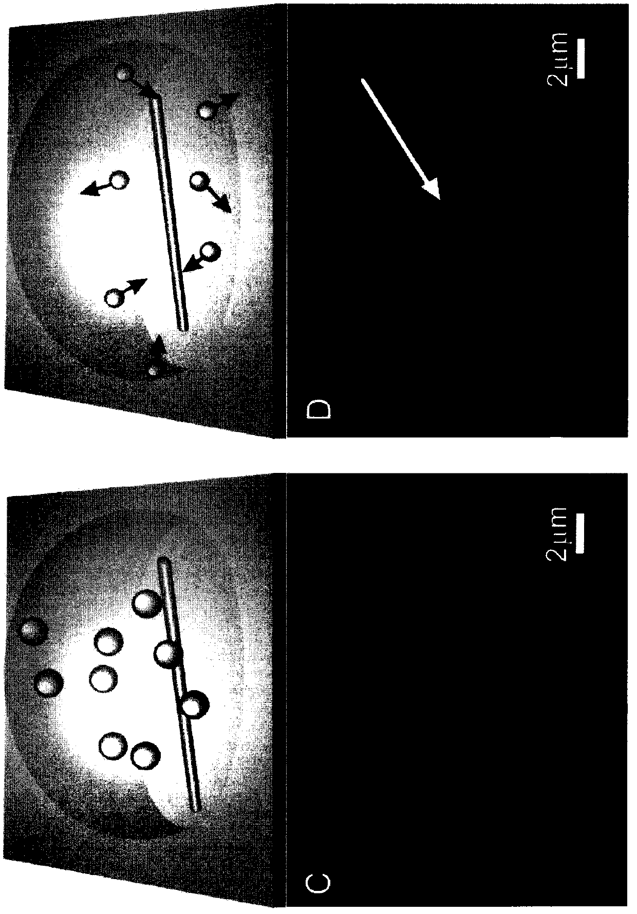

Image

  • A surface-enhanced Raman scattering substrate with visible hotspots, a preparation method, and a method for detecting molecules using the substrate
  • A surface-enhanced Raman scattering substrate with visible hotspots, a preparation method, and a method for detecting molecules using the substrate
  • A surface-enhanced Raman scattering substrate with visible hotspots, a preparation method, and a method for detecting molecules using the substrate

Examples


Embodiment 1

[0026] Step 1, preparation of silver nanowires

[0027] Add 10mL of ethylene glycol into a 100mL three-neck flask, heat it to reflux at 160°C for 2h, and inject 0.1M silver nitrate solution and 0.222g of polyvinylpyrrolidone (dissolved in 5mL of ethylene glycol solvent respectively) into it simultaneously with two syringe pumps. Middle), the injection rate was 0.2mL / min. Finally, react at a constant temperature of 160°C for 60 minutes. The synthesized silver nanowire sol is washed twice with acetone and secondary water according to the volume ratio of 1:9. The obtained silver nanowires have a length of 10 μm and a diameter of about 110 nm. .

[0028] Step 2, preparation of gold nanoparticles:

[0029] Add 90mL of secondary water and 1mL of 1% chloroauric acid solution into a 500mL three-neck flask, stir magnetically, heat the oil bath to boiling, then quickly add 1mL of 1% sodium citrate solution as a reducing agent , the diameter of the obtained gold nanoparticles is about...

Embodiment 2

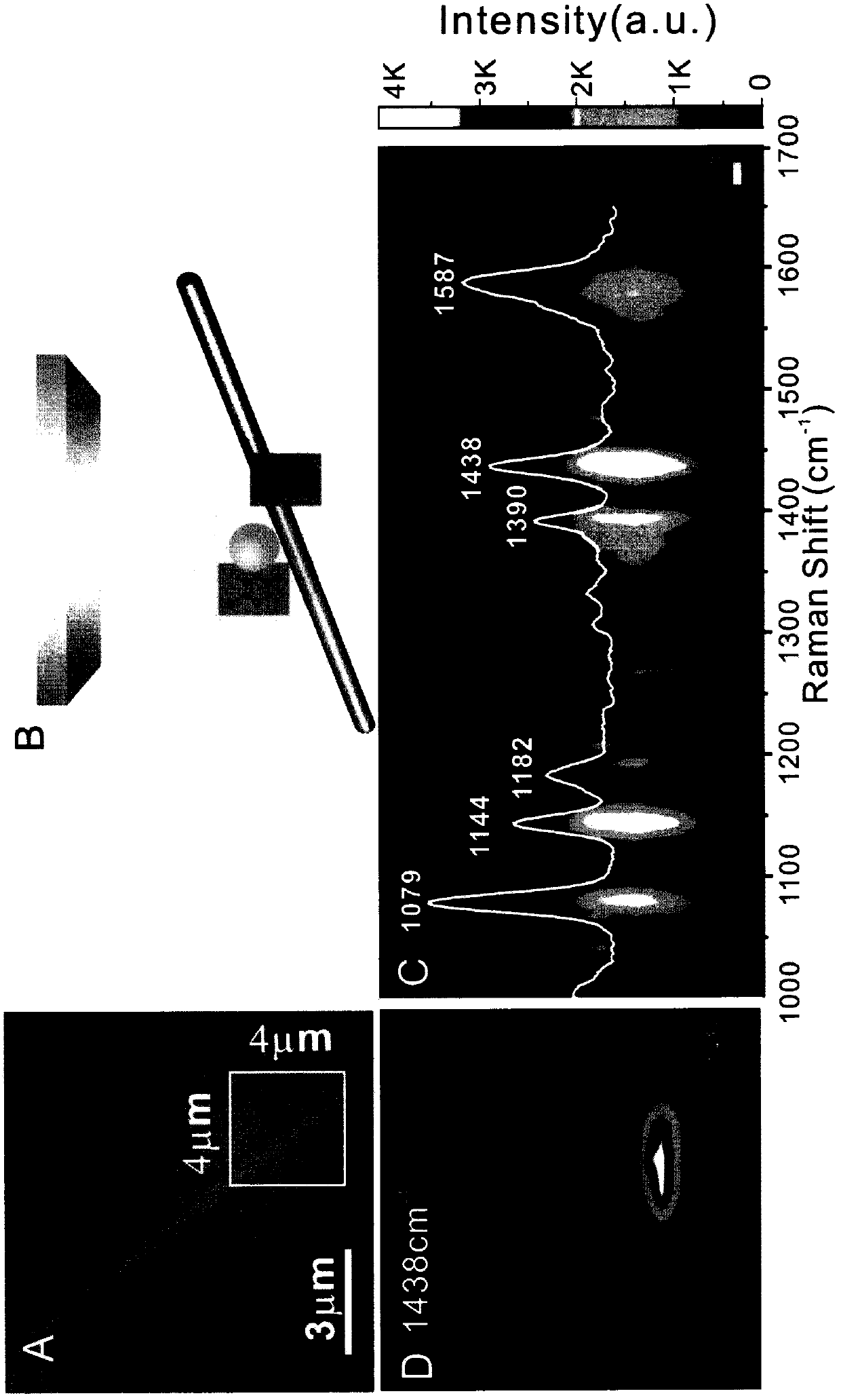
[0035] Use the SERS substrate prepared by the present invention to measure 4-ATP molecules, and obtain SERS spectra and regional SERSmapping images:

[0036] Place the SERS substrate synthesized in Example 1 under a Raman optical microscope, find the position where the silver nanowire surface is modified with gold nanoparticles, that is, the position where the SERS "hot spot" is generated, and pipette 1 μL with a concentration of 10 -8 M's 4-ATP solution, slowly add dropwise to the "hot spot" or near it. After drying for about 1 minute, select the excitation light with a wavelength of 633nm and a laser power of 2mW, integrate for 1 second, detect the signal, and obtain the SERS spectrum of the 4-ATP molecule.

[0037] figure 2 A is the Raman optical microscope image of the SERS substrate prepared by the present invention, where the box marked is the spot range of the Raman laser: (4×4) square microns; figure 2 B is a schematic diagram when the substrate prepared by the pre...

Embodiment 3

[0040] Utilize the SERS substrate prepared by the present invention to measure cysteine, adenosine triphosphate, methamphetamine and paraoxon:

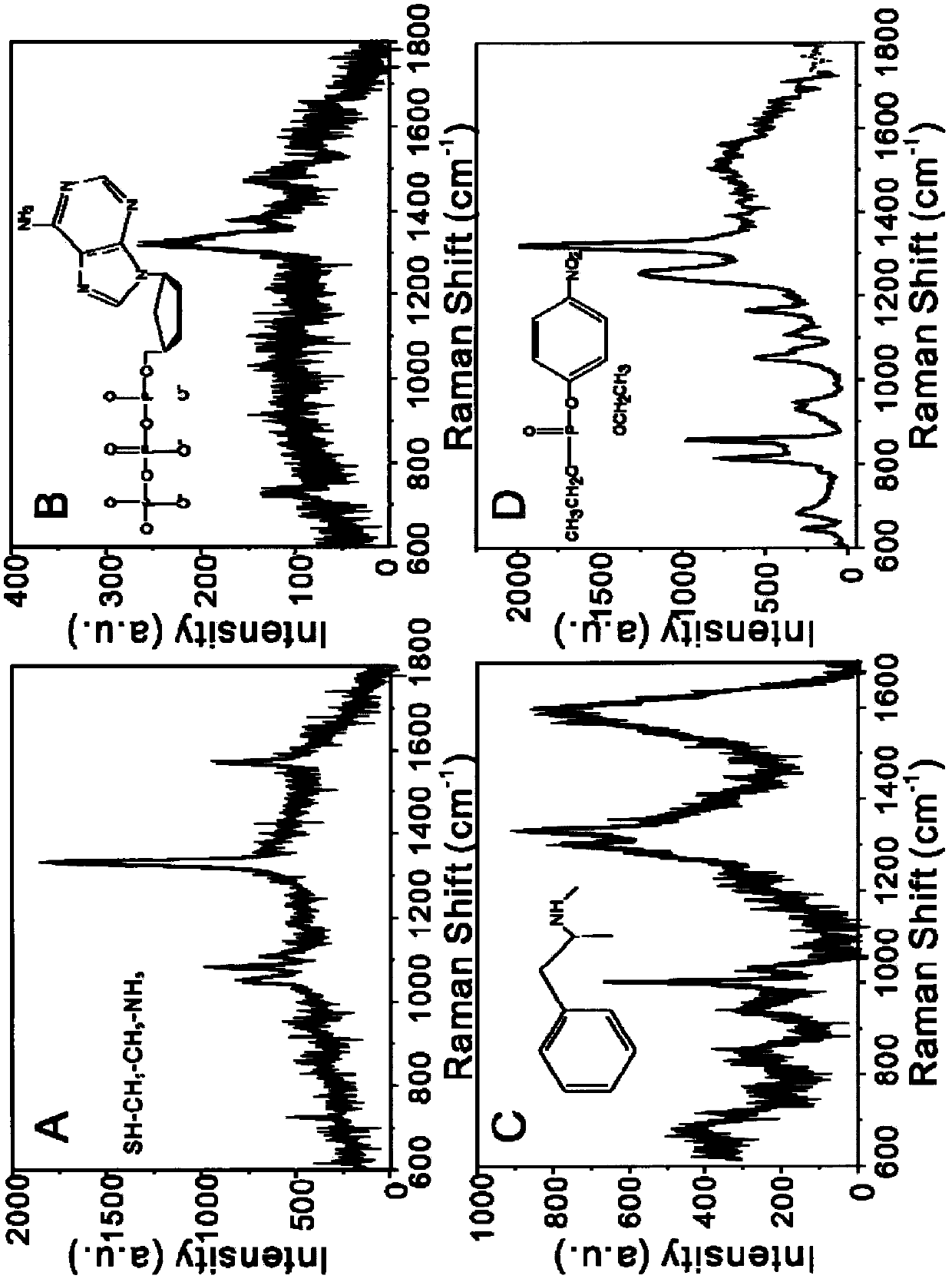
[0041] With the method identical with above-mentioned embodiment 2, can obtain respectively that concentration is 10 -6 M cysteine, 10 -6 M adenosine triphosphate, 10 -7 M of methamphetamine and 10 -8 SERS spectrum of paraoxon in M.

[0042] image 3 Medium, A: 10 -6 Cysteine ​​for M, B: 10 -6 ATP of M, C: 10 -7 Methamphetamine for M, D: 10 -8 M paraoxon, the results show that whether it is common cysteine ​​and adenosine triphosphate molecules in the human body, or common methamphetamine molecules and pesticide paraoxon molecules, the SERS substrate prepared by the present invention can achieve high-sensitivity SERS Detection effect.


PUM

PropertyMeasurementUnit
lengthaaaaaaaaaa
diameteraaaaaaaaaa
lengthaaaaaaaaaa

Abstract

The invention relates to a surface-enhanced Raman scattering substrate with visible hot spots, a preparation method and a method for detecting molecules by using the substrate. The SERS substrate is composed of silver nanowires placed on a Raman scattering substrate and gold nanoparticles modified on the surface of the silver nanowires. Plasmon resonance is formed between the silver nanowires and gold nanoparticles on the substrate, generating a strong electromagnetic field. Visible hot spots form. Place the SERS substrate under the Raman optical microscope, find the position of the gold nanoparticles modified on the surface of the silver nanowires, which is the SERS hotspot, and slowly add the molecules to be tested to this position, so that the molecules to be tested can effectively fall into the SERS hotspots within range. The preparation method simply utilizes capillary force and gravity to decorate gold nanoparticles on the surface of silver nanowires. The prepared substrate SERS has "visible hotspots", which can effectively make the molecules to be tested fall into the range of "hotspots", which will directly improve the sensitivity of the detection results.

Description

technical field [0001] The invention relates to a surface-enhanced Raman scattering technology and a related detection method, in particular to a surface-enhanced Raman scattering substrate with visible hotspots, a preparation method and a method for detecting molecules using the substrate. Background technique [0002] Since Martin Fleischmann and his collaborators discovered the surface-enhanced Raman scattering (SERS) phenomenon in 1974, and Jeanmaire and Van Duyne gave a theoretical explanation of this amazing discovery in 1977, SERS technology has been developed for nearly 40 years. years of history. SERS technology, because it provides rich fingerprint information of molecules, can distinguish the structural differences of isomers, can distinguish the different orientations of the same molecule to be tested on the SERS substrate, and realizes highly sensitive detection, so it has been widely studied and Application, is an advanced analytical detection method. [0003...

Claims


Application Information

Patent Timeline
Patent Type & Authority Patents(China)
IPC IPC(8): G01N21/65B22F9/24
Inventor 吴义平李盼杨良保
Owner HEFEI UNIV
Learn More