Double-antibody sandwich ELISA kit for detecting E-type enterovirus pathogen

An enterovirus, enzyme-labeled antibody technology, applied in the biological field, to achieve the effect of improving sensitivity and specificity, easy to use and strong specificity

Inactive Publication Date: 2016-05-04
JILIN UNIV
View PDF0 Cites 4 Cited by
  • Summary
  • Abstract
  • Description
  • Claims
  • Application Information

AI Technical Summary

Problems solved by technology

There are few reports on the diagnostic methods of BEV infection, especially the methods and kits for detecting entero

Method used


Image

  • Double-antibody sandwich ELISA kit for detecting E-type enterovirus pathogen
  • Double-antibody sandwich ELISA kit for detecting E-type enterovirus pathogen
  • Double-antibody sandwich ELISA kit for detecting E-type enterovirus pathogen

Examples


Embodiment 1

[0022] The construction of embodiment 1E enterovirus structural protein VP1 and VP2 prokaryotic expression recombinant plasmid

[0023] 1. Primer design

[0024] According to the base composition of the target sequence, primers were designed to amplify the VP1 and VP2 genes, and the upstream and downstream contained BamHI and EcoRI restriction sites respectively. The primer sequences are as follows:

[0025] Primers for amplifying the VP1 sequence:

[0026] E-VP1-FCGC GGATCC GAAACAAGCGTGGAGA (BamHI)

[0027] E-VP1-RCCG GAATTC GTACGAGGTGAGGCT (EcoRI)

[0028] Primers for amplifying the VP2 sequence:

[0029] E-VP2-FCGC GGATCC TCTCCGTCAGCAGAAG (BamHI)

[0030] E-VP2-RCCG GAATTC TGATGCAATAGCCCG (EcoRI)

[0031] 2. Gene amplification

[0032] Genomic RNA of Enterovirus E was extracted by the conventional Trizol method, and cDNA was obtained by using a commercially available conventional reverse transcription kit according to the instructions, and the VP1 gene and VP...

Embodiment 2V

[0037] Expression and purification of embodiment 2VP1 and VP2 recombinant protein

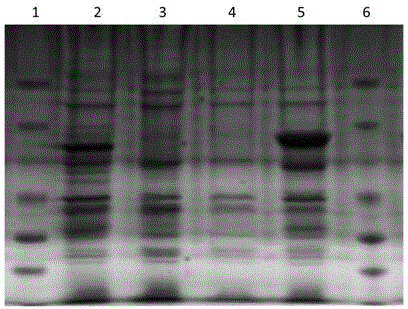
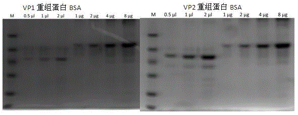
[0038] After transforming the identified positive recombinant plasmids pGEX-4T-1-VP1 and pGEX-4T-1-VP2 into BL21(DE3) competent cells, pick a single colony and inoculate it into 3 mL of LB liquid medium containing 100 μg / mL ampicillin culture overnight, then inoculated 1 mL of the above culture into 200 mL of LB liquid medium containing 100 μg / mL ampicillin and cultured with shaking at 37 °C until the logarithmic growth phase (OD 600 =0.6~0.8), adding IPTG to a final concentration of 1mmol / L, inducing culture at 20°C for 3h, and detecting by SDS-PAGE, the recombinant target proteins VP1 and VP2 were obtained, as shown in figure 2 shown. Centrifuge and precipitate the bacteria, and after sonication, use urea to purify inclusion bodies to obtain high-purity recombinant proteins, such as image 3 shown.

Embodiment 3

[0039] Example 3 Preparation and Purification of Anti-VP1 Mouse Polyclonal Antibody and VP2 Monoclonal Antibody

[0040] 1. Preparation of anti-VP1 mouse-derived polyclonal antibody: select healthy BALB / C mice aged 6-8 weeks, emulsify and purify recombinant VP1 protein with complete Freund's adjuvant, and inject 100 μg subcutaneously at multiple points in each mouse, and then On the 14th day, the same method was used to boost the immunization once, and the serum was collected at the same time to test the titer. For the last booster immunization, 100 μg of purified protein was directly injected intraperitoneally, and blood was collected 3 to 5 days later to collect serum, which was the anti-VP1 mouse polyclonal antibody;

[0041]2. Preparation of anti-VP2 monoclonal antibody: The immunization method is the same as that of anti-VP1 murine polyclonal antibody preparation. Take splenocytes from immunized mice and mix them with SP2 / 0 in a fusion tube, centrifuge at 300g for 10min, ...


PUM


Abstract

The invention discloses a double-antibody sandwich ELISA kit for detecting an E-type enterovirus pathogen. VP1 protein expressed by a VP1 gene with the base sequence shown as a sequence table SEQ ID NO.1 is adopted to immunize BALB/C mice to prepare a polyclonal antibody serving as a capture antibody, VP2 protein expressed by a VP2 gene with the base sequence shown as a sequence table SEQ ID NO.3 is adopted to immunize BALB/C mice to prepare a monoclonal antibody, and horse radish peroxidase (HRP) is marked. Safety is high, and the coincidence rate between kit detection results and RT-PCR detection results is 100%. The kit can be stored for 6 months at the temperature of 4 DEG C, and no obvious difference exists between detection results and those of a conventional method.

Description

technical field [0001] The invention belongs to the field of biological technology, in particular to a double-antibody sandwich ELISA kit for detecting E enterovirus pathogens. Background technique [0002] Bovine enterovirus (BEV) and human poliovirus, human Coxsackie virus, human intestinal cytopathic orphan virus, and porcine enterovirus are members of the genus Enterovirus in the family Picornaviridae. The resulting infectious diseases cause severe economic losses to the cattle industry. Clinically, bovine enterovirus infection is characterized by fever, lacrimation, cough, runny nose, dyspnea and severe diarrhea. Since the disease was first reported by Moll et al. in the 1950s, other countries have also reported the occurrence of this disease. In 2011, Li Yingli et al. first isolated a strain of enterovirus F from calf feces samples from a dairy farm in Inner Mongolia, confirming the existence of BEV infection in domestic cattle herds. In 2013, Peng Xiaowei and other...

Claims


Application Information

Patent Timeline
IPC IPC(8): C07K16/10G01N33/569G01N33/535
CPCC07K16/1009G01N33/535G01N33/56983G01N2333/085
Inventor 王新平邢泽黎朱利塞郭昌明盖小春王明月鲁海冰曹玉峰刘亚静张群
Owner JILIN UNIV
Why Eureka
Try Eureka