Ultrasonic contrast agent of uterus oviduct tract and preparation method of ultrasonic contrast agent

A technology for ultrasound contrast agents and fallopian tubes, which is applied in the field of medical contrast agents and injectable ultrasound targeting contrast agents. It can solve the problems of unsatisfactory particle size and distribution of microbubbles, complex preparation process, and complex synthesis process, etc., and achieve good clinical application. Foreground, uniform particle size distribution, and good biocompatibility

Active Publication Date: 2016-06-15
河北艾圣科技有限公司
View PDF3 Cites 5 Cited by
  • Summary
  • Abstract
  • Description
  • Claims
  • Application Information

AI Technical Summary

Problems solved by technology

This technology has achieved satisfactory results in diagnosing lesions in the ovary, intrauterine cavity, and fallopian tubes. However, there are some problems with the existing ultrasound contrast agents at home and abroad: for example, the ultrasonic microbubble membrane material has complex components, many materials, and synthetic preparation. In the process, chloroform, tert-butanol and other harmful solvents are used, so it is necessary to select the membrane material and its configuration ratio to prepare more economical, effective, safe and easy-to-prepare microbubbles; the synthesis process is complex and the steps are cumbersome , Ultrasonic cavitation method, mechanical formulation method, freeze-drying method, repeated freeze-thawing method, etc., either the preparation process is more complicated, or the particle size and distribution of the prepared microbubbles are not ideal enough, all of which need further improvement; Too many liposomes in the blood vessel or cavity can not release the gas in time, thus affecting the effect of ultrasound

Method used


Image

  • Ultrasonic contrast agent of uterus oviduct tract and preparation method of ultrasonic contrast agent
  • Ultrasonic contrast agent of uterus oviduct tract and preparation method of ultrasonic contrast agent
  • Ultrasonic contrast agent of uterus oviduct tract and preparation method of ultrasonic contrast agent

Examples


Embodiment 1

[0047] Dissolve 48 mg of lecithin, 2.4 mg of diarachidoylphosphatidylcholine (DAPC), 500 mg of polyethylene glycol-2000, 7.2 mg of poloxamer, and 600 mg of folic acid-polyethylene glycol phospholipid in 10 mL Place the mixture in a beaker of normal saline, then place the mixture in a water bath at 50°C, and stir it magnetically for 30 minutes to obtain a uniformly mixed solution;

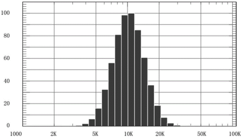
[0048] Will SF 6 The gas is passed into the solution, and then treated with a high-speed liquid shearing machine at 18000 rpm. At this time, the gas-encapsulated microsphere suspension is obtained. (Respectively at 13000rpm, 15000rpm, repeat the test)

Embodiment 2

[0050] Dissolve 48 mg of lecithin, 2.4 mg of diarachidoylphosphatidylcholine (DAPC), 500 mg of polyethylene glycol-2000, 7.2 mg of poloxamer, and 600 mg of folic acid-polyethylene glycol phospholipid in 10 mL Put the mixed solution in a beaker of normal saline, and then magnetically stir the mixed solution for 30 minutes in a water bath at 50°C to obtain a uniformly mixed solution; liposome suspension.

[0051] Leave the liposome suspension at room temperature for 24 hours, discard the supernatant, add an equal amount of distilled water to wash, purify 3 times, then discard the supernatant to obtain the upper layer mixture; vacuum freeze-dry the mixture Next, the liposome dry powder was obtained.

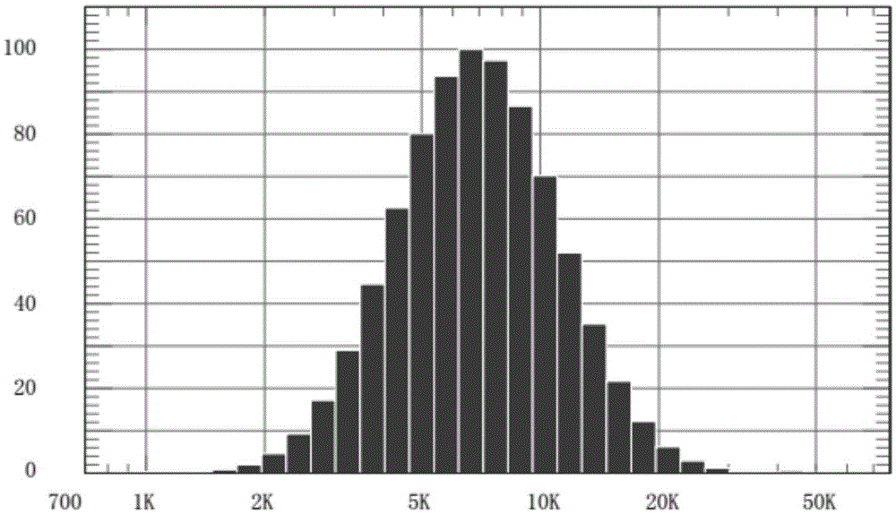
[0052] then SF 6 The gas is injected into the liposome dry powder, and then the dry powder obtained at this time is dissolved in physiological saline to obtain a stable ultrasound contrast agent with uniform particle size.

Embodiment 3

[0054] Dissolve 48 mg of lecithin, 2.4 mg of diarachidoylphosphatidylcholine (DAPC), 500 mg of polyethylene glycol-2000, 7.2 mg of poloxamer, and 600 mg of folic acid-polyethylene glycol phospholipid in 10 mL Place the mixture in a beaker of normal saline, then place the mixture in a water bath at 50°C, and stir it magnetically for 30 minutes to obtain a uniformly mixed solution;

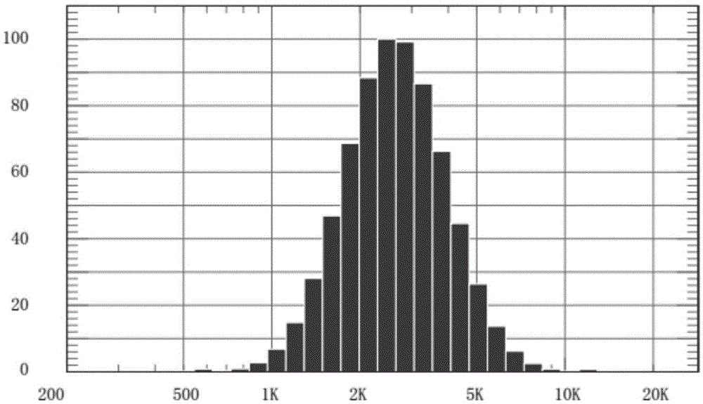
[0055] Pass perfluoropentane into the solution, and then process it with a high-speed liquid shearing machine at 18000 rpm, at this time, a suspension of microspheres wrapped in perfluoropentane is obtained.


PUM


Abstract

The invention relates to an ultrasonic contrast agent of a uterus oviduct tract and a preparation method of the ultrasonic contrast agent. The ultrasonic contrast agent is prepared from the following components: a first component is a carrier and is composed of phospholipid, polyethylene glycol or a derivative thereof and polymerized phospholipid connected with a specific ligand at the weight ratio of (0.5 to 2):(0.005 to 0.02):(0.003 to 0.007); a second component is a phospholipid stabilizer; a third component is a biological solvent; a fourth component is a fluorocarbon gas; the weight percent ratio of the first component to the second component to the third component is (0.0005 to 0.002):(0.000005 to 0.00002):(0.5 to 2); the concentration of the fourth component which is gas is 10<8>-10<9> / mL.

Description

technical field [0001] The invention relates to a medical contrast agent, in particular to an injectable ultrasound-targeted contrast agent with air-filled microbubbles in an aqueous solution carrier, belonging to the field of medical ultrasound image diagnosis. Background technique [0002] Suspensions of gaseous microbubbles in a carrier liquid act as effective ultrasound reflectors and are well known in the art. With the application of ultrasound, although transvaginal ultrasound has a clearer image of female internal genitalia than the previous abdominal ultrasound, general real-time ultrasound still cannot fully display the course and shape of the fallopian tubes. Or ovarian lesions, can not be clearly displayed. At present, hysterosalpingosalpingography and laparoscopy are commonly used at home and abroad to check the patency of the fallopian tubes, but many patients often have allergic reactions to lipiodol, and the entry of lipiodol contrast agents into the abdomina...

Claims


Application Information

Patent Timeline
Patent Type & Authority Applications(China)
IPC IPC(8): A61K49/22
CPCA61K49/221A61K49/223
Inventor 袁哲杨赣英
Owner 河北艾圣科技有限公司
Why Eureka
Try Eureka