Liquid-phase suspension biochip based on multi-optical trap encoding bead array and two-photon fluorescence detection

A biochip, coding microsphere technology, applied in fluorescence/phosphorescence, measurement devices, material analysis by optical means, etc., can solve the problem of limited number of optical traps, and achieve the simplification of enrichment process, cost reduction, and simplification of structure. Effect

Inactive Publication Date: 2016-07-20
WUHAN UNIV
View PDF5 Cites 37 Cited by
  • Summary
  • Abstract
  • Description
  • Claims
  • Application Information

AI Technical Summary

Problems solved by technology

The time-sharing scanning optical tweezers technology controls the deflection of the laser through scanning galvanometers (GM), acousto-optic deflectors (AOD) and other devices, so that a single laser beam can scan quickly on the focal plane. The particles can be stably bound on the laser scanning path when the Brownian motion time of the particle is greater than its minimum residence time, and the particle’s Brownian motion time is longer than its departure time. Although the time-sharing scanning technology does not require high laser power, theoretically, this method can Generates a limited number of optical traps (typically less than 100)

Method used


Image

  • Liquid-phase suspension biochip based on multi-optical trap encoding bead array and two-photon fluorescence detection
  • Liquid-phase suspension biochip based on multi-optical trap encoding bead array and two-photon fluorescence detection
  • Liquid-phase suspension biochip based on multi-optical trap encoding bead array and two-photon fluorescence detection

Examples


Embodiment 1

[0048] Detection of five liver cancer markers AFP, CEA, Glypican-3 (GPC-3), abnormal prothrombin (DCP) and α-L-fucosidase (AFU)

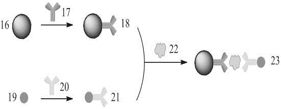
[0049] Five 3μm colored microspheres (blue, green, yellow, red, colorless) with surface modified carboxyl groups were used as color-coded microspheres, and AFP, CEA, GPC-3, DCP were enriched by two-site sandwich method. , AFU antigen, and use quantum dots with emission wavelength of 605nm as fluorescent labeling materials, such as image 3 Shown.

[0050] The first step is to prepare immuno-encoded microspheres. Take one of the blue coded microspheres 16 modified with surface carboxyl groups, first activate the carboxyl groups on the surface of the microspheres through the EDC / NHS reaction, and then couple one of the AFP monoclonal antibodies 17 at room temperature to obtain immunomicrospheres 18. The preparation methods of the remaining four immuno-encoding microspheres are similar, and finally the five immuno-encoding microspheres are mixed into a buf...

Embodiment 2

[0056] Quantitative detection of H1N1, H3N2, H9N2 three avian influenza viruses

[0057] Using size coding method, the surface modified polystyrene microspheres with particle size of 1μm, 2μm, and 3μm were used as solid-phase carriers, and H1N1, H3N2, H9N2 viruses were enriched by the two-site sandwich method, and emitted Quantum dots with a wavelength of 605nm are used as fluorescent labeling materials, such as Figure 4 Shown.

[0058] The first step is to prepare immuno-encoded microspheres. Take one of the coded microspheres 24 with a surface carboxyl group modification and a particle size of 1 μm. The carboxyl groups on the surface of the microspheres are first activated by the EDC / NHS reaction, and then one of the H1N1 monoclonal antibodies 25 is coupled at room temperature to obtain an immunomicrobe Ball 26, the preparation methods of the other two immunomicrospheres are similar, and finally the three immunoencoding microspheres are mixed into the buffer solution for use. ...


PUM

PropertyMeasurementUnit
particle diameteraaaaaaaaaa

Abstract

The invention provides a liquid-phase suspension biochip based on multi-optical trap encoding bead array and two-photon fluorescence detection. The liquid-phase suspension biochip has a configuration as follows: a near-infrared laser beam emitted by a near-infrared laser is expanded by a beam expanding system, is sequentially reflected by a telescope system and a dichroscope by using a holographic technology or a time-share scanning technology, and is focused into a sample pool through a high numerical aperture objective lens, to form multi-optical-trap optical tweezers; the multi-optical-trap optical tweezers capture a plurality of encoding beads enriched with objects to be detected, to form a bead array in the solution; after an infrared laser signal is filtered by a band-pass filter, a two-photon fluorescence signal from each bead is focused to an image detector by a lens and is subjected to imaging detection. The liquid-phase suspension biochip can perform real-time quantitative analysis on nucleic acids, proteins, virus particles and a plurality of objects to be detected, and has the advantages of high sensitivity, strong anti-interference ability, simultaneous determination of a plurality of components and the like.

Description

Technical field [0001] The invention belongs to the technical field of optical trap optical tweezers micromanipulation, and also belongs to the technical field of microsphere encoding, and specifically relates to a liquid-phase suspension type biological chip analysis system based on a multi-light trap encoding microsphere array and two-photon fluorescence detection. Background technique [0002] Traditional solid-phase biochips integrate the biochemical analysis process on the surface of solid-phase transmitters such as silicon wafers, glass sheets, nylon membranes, etc., based on the principle of specific interactions between biomolecules, to achieve high-throughput rapid detection of multiple biological components . This technology is widely used, but because the reaction is carried out on the surface of the solid phase, it has defects such as low reaction efficiency, poor stability and reliability. In the late 1990s, American Luminex company developed a multifunctional liqui...

Claims


Application Information

Patent Timeline
Patent Type & Authority Applications(China)
IPC IPC(8): G01N21/64
CPCG01N21/6486G01N21/6458
Inventor 唐宏武李诚予曹迪康亚峰庞代文
Owner WUHAN UNIV
Why Eureka
Try Eureka