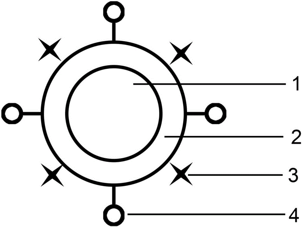
Preparation method of fluorogold nanocomposite used for cell imaging
A fluorescent gold nanometer and cell imaging technology, which is applied in the field of preparation of fluorescent gold nanocomposites, can solve the problems of easy photobleaching, limit the application of fluorescence methods, and low brightness, and achieve broad application prospects, increase fluorescence intensity, and low cost. Effect
- Summary
- Abstract
- Description
- Claims
- Application Information
AI Technical Summary
Problems solved by technology
Method used
Image
Examples
preparation example Construction
[0021] An embodiment of the present invention is a method for preparing a fluorescent gold nanocomposite for cell imaging, comprising:
[0022] The step of synthesizing gold nanoparticles; the step of coating the polylysine shell on the surface of the gold nanoparticles; and using the reaction between the amino group and the succinimide group on the surface of the shell to modify the interaction between the fluorescent dye and the targeting molecule step.
[0023] Preferably, the step of synthesizing gold nanoparticles is: soak the flask with aqua regia for 30 minutes, and clean it with deionized water; add deionized water and amino acid respectively in the ratio of adding 5 mg of amino acid to every 10 mL of deionized water in the flask, at room temperature Stir fully to dissolve; then add chloroauric acid solution in a ratio of 100 μL for every 5 mg of amino acid, the concentration of the chloroauric acid solution is 0.1mol / L, stir at room temperature for 3 hours, and the so...
Embodiment 1
[0034] A method for preparing a fluorescent gold nanocomposite for cell imaging, comprising the following steps:
[0035] Step 1. Synthesis of gold nanoparticles
[0036] Take a 25mL round-bottom flask, soak it in aqua regia for 30 minutes, and then clean it with deionized water. Add 10 mL of deionized water and 5 mg of tryptophan into the flask, stir well at room temperature to dissolve. Then add 100 μL chloroauric acid solution (0.1mol / L concentration), stir at room temperature for 3 hours, the solution gradually changes from colorless to dark red;
[0037] Step 2, coating of polylysine shell
[0038]Dilute 2 mL of the gold nanoparticle solution prepared in step 1 to 4 mL and add it to a 10 mL round bottom flask. Add 100 μL of polylysine solution (concentration: 25 mg / mL) and 0.2 mg of disuccinimide suberate to it, adjust pH=9, and stir at room temperature for 3 hours;
[0039] Step 3. Modification of fluorescent dyes and targeting molecules
[0040] Centrifuge the coat...
Embodiment 2
[0042] A method for preparing a fluorescent gold nanocomposite for cell imaging, comprising the following steps:
[0043] Step 1. Synthesis of gold nanoparticles
[0044] Take a 25mL round-bottomed flask, soak it in aqua regia for 30 minutes and then clean it with deionized water; add 10mL deionized water and 5mg tryptophan to the flask, and stir well at room temperature to dissolve. Then add 100 μL chloroauric acid solution (0.1mol / L), stir at room temperature for 3 hours, the solution gradually changes from colorless to dark red;
[0045] Step 2, coating of polylysine shell
[0046] Dilute 2 mL of the gold nanoparticle solution prepared in step 1 to 4 mL and add it to a 10 mL round bottom flask. Add 100 μL of polylysine solution (25 mg / mL) and 0.2 mg of 3,3’-dithiobis(sulfosuccinimidyl propionate) to it, adjust the pH to 9, and stir at room temperature for 3 hours;
[0047] Step 3. Modification of fluorescent dyes and targeting molecules
[0048] Centrifuge the coated pr...
PUM
| Property | Measurement | Unit |
|---|---|---|
| concentration | aaaaa | aaaaa |
Abstract
Description
Claims
Application Information
 - R&D
- Intellectual Property
- Life Sciences
- Materials
- Tech Scout
- Unparalleled Data Quality
- Higher Quality Content
- 60% Fewer Hallucinations
Browse by: Latest US Patents, China's latest patents, Technical Efficacy Thesaurus, Application Domain, Technology Topic, Popular Technical Reports.
© 2025 PatSnap. All rights reserved.Legal|Privacy policy|Modern Slavery Act Transparency Statement|Sitemap|About US| Contact US: help@patsnap.com

