Live animal two-photon excitation time-lapse detection fluorescence imaging analysis method and equipment
An analysis method, a technique of fluorescence imaging, which is applied in the field of in vivo fluorescence image analysis of animals, and can solve problems such as difficult application of high-efficiency two-photon excitation imaging analysis
- Summary
- Abstract
- Description
- Claims
- Application Information
AI Technical Summary
Problems solved by technology
Method used
Image
Examples
Embodiment 1
[0069] Example 1. Analysis of tumor targeting kinetics of fluorescent nanoprobes in tumor-bearing mice
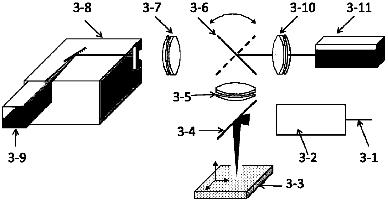
[0070] The HepG-2 liver cancer model for two-photon excitation time-resolved imaging was established on the right hind leg of nude mice. In the experiment, transferrin (Tf) and RGD modified with 10% Eu(tta) were used respectively. 3 Styrene-maleic anhydride copolymer (SMA) nanoparticles (16nm) of bpt obtained two photon-sensitized Eu 3+ Luminescent fluorescent nanoprobes. 300 microliters of colloid solution containing 1.5 mg of fluorescent nanoprobes was injected through the tail vein, and then the mice were anesthetized using an inhalation respiratory anesthesia system to ensure that the mice would not produce significant displacement during the imaging process. On the two-photon excitation time-resolved imaging device of the present invention, a femtosecond pulsed laser with a wavelength of 800nm is used to excite the fluorescent nanoprobe in the body of the desired a...
Embodiment 2
[0074] Example 2. Comparison of two-photon excitation time-lapse fluorescence detection imaging and ordinary two-photon excitation fluorescence imaging in live animal imaging analysis
[0075] In this embodiment, the conditions of the two-photon excitation time-lapse detection imaging experiment are the same as those in Embodiment 1, and the excitation conditions are the same in the ordinary two-photon excitation fluorescence imaging experiment, and the time delay is zero nanoseconds. We performed two-photon excitation time-lapse fluorescence detection imaging and ordinary two-photon excitation fluorescence imaging on the tumor area of tumor-bearing mice before and after fluorescent probe injection. The result is as Figure 8 shown. The experimental results show that using the method and device provided by the present invention can effectively eliminate the influence of background fluorescence on imaging analysis in two-photon excitation live animal imaging analysis, and ob...
Embodiment 3
[0076] Embodiment 3, the detection depth experiment of the imaging analysis method of the present invention
[0077] Fix the fluorescent nanoprobe on the bottom of the container, inject the adipose tissue simulation liquid into the container, adjust the thickness of the adipose tissue simulation liquid covered on the bottom probe, and use the time-lapse fluorescence detection method and equipment of the present invention to allow the excitation light to pass through the top The probe is excited by the tissue simulating fluid, and imaging analysis is performed above the liquid surface. The excitation and detection conditions are as in Example 1. Such as Figure 9 As shown, the results show that the detection depth of fluorescence imaging in this experiment is greater than 7mm, which is significantly improved compared with the previous reliable detection depth of fluorescence imaging. Studies have shown that the detection depth can be further greatly improved by changing the ex...
PUM
| Property | Measurement | Unit |
|---|---|---|
| size | aaaaa | aaaaa |
| size | aaaaa | aaaaa |
| wavelength | aaaaa | aaaaa |
Abstract
Description
Claims
Application Information
 - R&D
- Intellectual Property
- Life Sciences
- Materials
- Tech Scout
- Unparalleled Data Quality
- Higher Quality Content
- 60% Fewer Hallucinations
Browse by: Latest US Patents, China's latest patents, Technical Efficacy Thesaurus, Application Domain, Technology Topic, Popular Technical Reports.
© 2025 PatSnap. All rights reserved.Legal|Privacy policy|Modern Slavery Act Transparency Statement|Sitemap|About US| Contact US: help@patsnap.com


