Lipidosome medicine carrying method

A liposome and blank liposome technology, applied in the field of biomedicine, can solve the problems of phospholipid decomposition, deterioration, lack of drug-carrying methods, etc., and achieve the effects of high stability and wide application range

Inactive Publication Date: 2016-09-21
ANHUI MEDICAL UNIV
View PDF3 Cites 4 Cited by
  • Summary
  • Abstract
  • Description
  • Claims
  • Application Information

AI Technical Summary

Problems solved by technology

Obviously, the ion gradient method has a limited scope of application, and is only suitable for partially dissociable amphoteric drugs or drugs that can complex with ions, and is not suitable for drugs that are sensitive to acids, bases, and metal ions; High or low pH values ​​can also easily cause the decomposition and deterioration of unstable phospholipids, and even lead to the disintegration of liposomes
Therefore, for most drugs, there is still a lack of effective drug-loading methods, and it is impossible to develop liposome preparations with clinical value.

Method used


Image

  • Lipidosome medicine carrying method

Examples


Embodiment 1

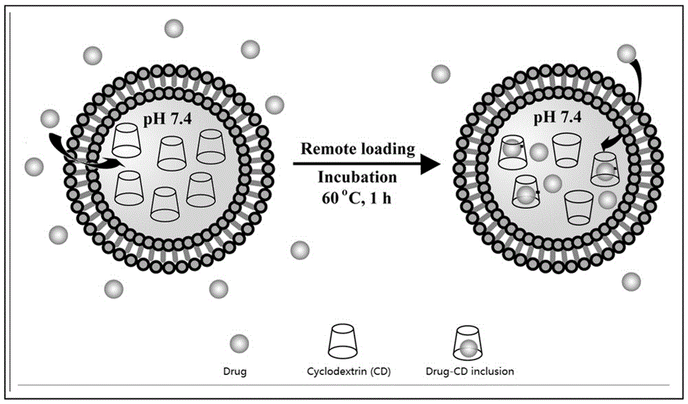
[0024] 300 mM α-cyclodextrin (α-CD) was used as the aqueous phase, and phosphatidylcholine and phosphatidylserine (SPC / SPS = 10:1, mole ratio) were used as membrane materials, and the package was prepared by emulsification-lyophilization method. Seal α-CD liposomes, remove free α-CD by centrifugation, and obtain blank liposomes encapsulating cyclodextrin (cyclodextrin: liposome membrane material = 1:5, mass ratio); then add baicalein, Drug / phospholipid (1:20, w / w), incubate at 40 °C for 1 hour to complete active drug loading; observe the liposome as a spherical nanoparticle by electron microscope, the average particle size detected by DLS is 220 nm, PDI = 0.36, ζ = At -30 mV, the encapsulation efficiencies detected by HLPC were 92%, 95%, and 99%, respectively. (SPC, soybean phosphatidylcholine; SPS, soybean phosphatidylserine; DLS, dynamic light scattering; PDI, polydispersity index).

Embodiment 2

[0026] Preparation of liposomes loaded with amphoteric drugs by CRL method

[0027] Using 100 mM PEG2000-hydroxypropyl-β-cyclodextrin (PEG-HP-β-CD) as the aqueous phase, phospholipids and cholesterol (DPPC / CHO = 1:1, mole ratio) as the membrane material, the package was prepared by the reverse evaporation method. Encapsulate HP-β-CD liposomes, remove free PEG-HP-β-CD by column chromatography, and obtain blank liposomes encapsulating cyclodextrin (cyclodextrin: liposome membrane material = 1:10, mass Ratio); then add Folate-PEG2000-DSPE (FPD) and topotecan (FPD / DPPC = 1:20, moleratio, drug / DPPC = 1:20, w / w), incubate at 45 °C for 1 hour, and complete the lipolysis Folic acid modification and active drug loading on the surface of plastids; the liposomes are spherical nanoparticles observed by electron microscopy, the average particle size detected by DLS is 300 nm, and the distribution is relatively uniform (PDI = 0.23), ζ = -5mV, (high performance liquid phase) HLPC The detect...

Embodiment 3

[0029] Preparation of oligopeptide-loaded vesicles by CRL method

[0030] 300 mM hydroxypropyl-β-cyclodextrin (HP-β-CD) and 5% sucrose were used as the aqueous phase, and SPAN 80 (SPAN80) and DOTAP (SPAN / DOTAP = 20:1, mole ratio) were used as membrane materials. Prepare positively charged vesicles encapsulating HP-β-CD by dispersion-extrusion method, remove free HP-β-CD by dialysis, and obtain blank liposomes encapsulating cyclodextrin (cyclodextrin: liposome membrane material = 1:15, mass ratio); then add glutathione (Glutathione / SPAN = 1:30, w / w) and incubate at 40 °C for 0.5 h to complete active drug loading; observe the liposomes as spherical nanoparticles by electron microscope , the average particle size detected by DLS is 200 nm, the distribution is uniform (PDI=0.23), ζ = 35mV, and the encapsulation efficiency detected by HLPC is 75%. The drug-loaded vesicles were further freeze-dried, and the freeze-dried product was stored at 4 °C for 6 months. After hydration, the ...


PUM

PropertyMeasurementUnit
particle diameteraaaaaaaaaa
particle sizeaaaaaaaaaa

Abstract

The invention relates to a lipidosome medicine carrying method. The method comprises the steps of 1, preparing cyclodextrin-encapsulated blank lipidosome, wherein the cyclodextrin-encapsulated blank lipidosome is lipidosome in which medicine to be encapsulated is not encapsulated but cyclodextrin is encapsulated, and the mass ratio of cyclodextrin to a lipidosome membrane material is 1:(5-15); 2, removing free cyclodextrin; 3, obtaining medicine-carrying lipidosome after co-incubation of the medicine to be encapsulated and the cyclodextrin-encapsulated blank lipidosome. The method has all the advantages of an ion gradient method, and also has the advantages that application range is wide, and limitation of medicine dissociation or complexing property is avoided; stability is high, and the pH of solution is neutral, so that the stability of components easy to hydrolyze such as phospholipid can be maintained; cyclodextrin is a polysaccharide compound, and integrity of a phospholipid membrane during freeze-drying can be maintained easily.

Description

technical field [0001] The invention relates to the technical field of biomedicine, and is a new active drug loading method of liposomes. Background technique [0002] Liposomes are closed vesicles formed by phospholipid bilayers. Liposomes are divided into unilamellar vesicles, multilamellar vesicles and multivesicular liposomes according to their structure. Liposomes were first discovered by British Alec D. Bangham in the early 1960s. Since then, it has been found that liposomes have great application value as material carriers, especially drug carriers, and large-scale systematic research on liposomes has been carried out immediately. [0003] After more than 60 years of exploration, researchers have developed many valuable liposome preparation methods. At present, liposome preparation methods are mainly based on phospholipid dispersion technique (dispersion technique), which can be classified into the following three categories: (1) Based on solvent or cosolvent (coso...

Claims


Application Information

Patent Timeline
Patent Type & Authority Applications(China)
IPC IPC(8): A61K9/127A61K47/24A61K47/28A61K47/26A61K47/18A61K47/48
CPCA61K9/127A61K9/1273A61K9/1277A61K47/186A61K47/24A61K47/26A61K47/28
Inventor 王汀王宁
Owner ANHUI MEDICAL UNIV
Why Eureka
Try Eureka