Human body thoracic and abdominal cavity CT image aorta segmentation method based on GVF Snake model

A CT image, thoracic and abdominal technology, applied in the field of medical image processing, can solve the problems of human life-threatening, heavy workload, etc., and achieve the effect of avoiding uncertainty, improving segmentation accuracy, and good repeatability

Inactive Publication Date: 2016-09-28
TIANJIN POLYTECHNIC UNIV
View PDF3 Cites 19 Cited by
  • Summary
  • Abstract
  • Description
  • Claims
  • Application Information

AI Technical Summary

Problems solved by technology

[0002] For patients with cardiovascular and cerebrovascular diseases, thoracoabdominal aortic dissection (AoD) is much more life-threatening than cerebral infarction, myocardial infarction and malignant tumors. The dissection is peeled off, resulting in the appearance of true lumen and false lumen in the aorta in the human thoracoabdominal cavity. When the blood flows in the true and false lumen, the blood vessel wall is squeezed, and at the same time, the blood enters the middle layer of the aorta through the intima to form a hematoma. Patients do not fully understand the condition, so the mortality rate within 48 hours after the onset of the condition can be as high as 36% to 71%, which poses a great threat to human life; currently, the main treatment for aortic dissection is endovascular isolation. As for the chief surgeon, it is necessary to obtain as much information as possible about the spatial relationship between the local anatomy and adjacency of the lesion during the operation, so as to achieve accurate diagnosis, operation and postoperative evaluation, especially for the location and extent of thoracoabdominal aortic dissection. It is one of the main factors that determine the indications of surgery and the success of the operation; therefore, it is very necessary to develop a medical image processing system to quickly and accurately extract the thoracoabdominal aorta and its dissection It is of great significance to improve the success rate of the operation; because each patient has a large number of CT tomographic images, the workload of manual segmentation is very large. For the three-dimensional image This is especially true for operations; another problem is that there is uncertainty in manual segmentation, and there are great differences in the segmentation results of different medical experts, and even the segmentation results of the same image by the same expert at different times and in different states are also different. Not a small difference; if the CT image can be automatically segmented, then the problems associated with the manual segmentation method will be solved; currently the most clinically used 3D reconstruction based on CT tomographic images, except for a few tissues with obvious contrast between the Housfield value and the surrounding , such as bones, lungs, and blood vessels after angiography, other tissues and organs cannot be automatically segmented; even for these tissues, due to the influence of scanning layer thickness and volume effect, the contours of automatic computer segmentation are often unsatisfactory to medical researchers; Therefore, it is currently a difficult point to accurately and quickly extract the aorta from complex and irregular tissues and organs.

Method used


Image

  • Human body thoracic and abdominal cavity CT image aorta segmentation method based on GVF Snake model
  • Human body thoracic and abdominal cavity CT image aorta segmentation method based on GVF Snake model
  • Human body thoracic and abdominal cavity CT image aorta segmentation method based on GVF Snake model

Examples


Embodiment Construction

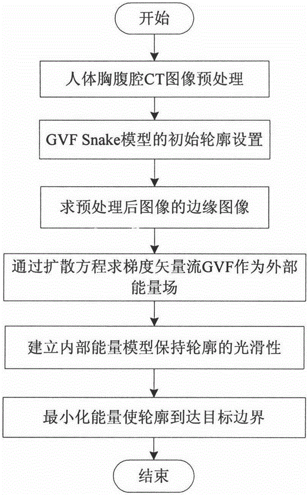
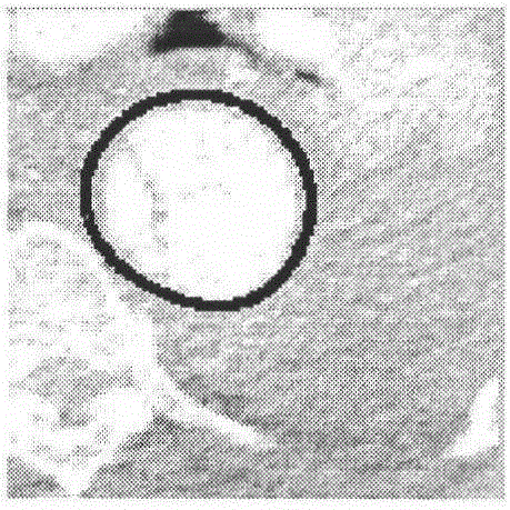
[0024] Algorithm flow chart of the present invention is as figure 1 As shown, first read the CT image and perform image preprocessing; then set the initial contour of the GVF Snake model on the preprocessed image; then obtain the edge image of the preprocessed image; based on the obtained edge image Then use the diffusion equation to find the gradient vector flow GVF as the external energy field; then establish the internal energy model to maintain the smoothness of the contour; finally use the internal energy and external energy to construct the energy function E, and obtain the minimum of the energy E through iterative operations value, and finally make the contour reach the target boundary. The specific implementation process of the technical solution of the present invention will be described in detail below in conjunction with the accompanying drawings.

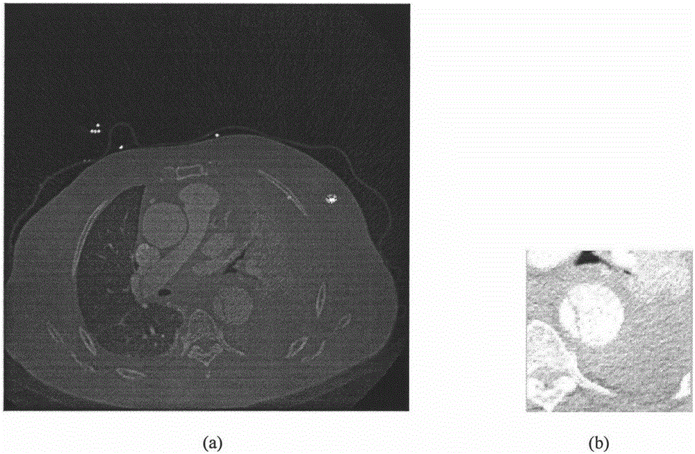
[0025] 1. Read CT images and perform image preprocessing

[0026] Due to the complexity of the internal structure of...


PUM


Abstract

The invention discloses a human body thoracic and abdominal cavity CT image aorta segmentation method based on a GVF Snake model. The method overcomes the shortcomings of the heavy workload and long time consuming of the traditional manual and semi-automatic segmentation, the repeatability of the method is good, and the uncertainty caused by artificial segmentation is prevented. The method includes (1) reading a CT image and performing image preprocessing; (2) performing the initial profile setting of the GVF Snake model on the image obtained after the preprocessing; (3) obtaining the edge image of the image after the preprocessing; (4) obtaining gradient vector flow GVF as the external energy field by the diffusion equation based on the obtained edge image; (5) establishing an internal energy model to maintain the smoothness of the profile; and (6) constructing an energy function E by means of internal energy and external energy, obtaining the minimum value of energy E by means of iteration operation, and the target boundary of the profile can be obtained at the end. The method has important application values in the field of human body thoracic and abdominal cavity aorta interlayer segmentation diagnosis treatment.

Description

technical field [0001] The invention belongs to the technical field of medical image processing, and relates to a GVF Snake model-based aorta segmentation method in a CT image of a human thoracoabdominal cavity, which can be used for three-dimensional reconstruction of the aorta in a human thoracoabdominal cavity. Background technique [0002] For patients with cardiovascular and cerebrovascular diseases, thoracoabdominal aortic dissection (AoD) is much more life-threatening than cerebral infarction, myocardial infarction and malignant tumors. The dissection is peeled off, resulting in the appearance of true lumen and false lumen in the aorta in the human thoracoabdominal cavity. When the blood flows in the true and false lumen, the blood vessel wall is squeezed, and at the same time, the blood enters the middle layer of the aorta through the intima to form a hematoma. Patients do not fully understand the condition, so the mortality rate within 48 hours after the onset of th...

Claims


Application Information

Patent Timeline
Patent Type & Authority Applications(China)
IPC IPC(8): G06T7/00
CPCG06T2207/10081G06T2207/20116
Inventor 段晓杰时美晨汪剑鸣陈丹丹赵鹤
Owner TIANJIN POLYTECHNIC UNIV
Why Eureka
Try Eureka