Vascularization support for tissue engineering and construction method of vascularization support
A tissue engineering scaffold, tissue engineering technology, applied in tissue regeneration, medical science, prosthesis, etc., can solve the problems of hindering cell migration, long angiogenesis time, blood flow disorder, etc., to shorten the vascularization time and shorten the vascularization. time, the effect of reducing the time to vascularization
- Summary
- Abstract
- Description
- Claims
- Application Information
AI Technical Summary
Problems solved by technology
Method used
Image

Examples
Embodiment 1
[0048] The tissue engineering vascularized stent of this embodiment is prepared by the following method:
[0049] (1) Preparation of porcine decellularized pericardium grafted with VEGF
[0050] Refer to patent CN101274106 B (a method for preparing acellular matrix) to prepare porcine decellularized pericardium, the specific steps are as follows: pretreat porcine pericardium tissue, and then add the pretreated porcine pericardium tissue to a mixture containing phospholipids The porcine decellularized pericardium is prepared by soaking in the enzyme solution through hypotonic or isotonic immersion, and then washed to obtain the porcine decellularized pericardium. Wherein, the concentration of phospholipase is less than 10000U / mL, the temperature range of the solution is 0-56°C, and the pH value range is 5-12.
[0051] Immerse 1 g of dry weight decellularized pericardium in 5 mL of 0.05 mol / L MES (2-(N-morpholine) ethanesulfonic acid monohydrate) buffer (pH=5.60) for 0.5 h; EDC...
Embodiment 2
[0072] The tissue engineering vascularized stent of this embodiment is prepared by the following method:
[0073] (1) Preparation of dog decellularized bladder loaded with strontium ions
[0074] Refer to patent CN 101274106 B (a method for preparing acellular matrix) to prepare dog decellularized bladder, then immerse 2g of dry weight dog decellularized bladder into 50mL of 0.05mol / L strontium chloride in PBS (pH=7.2) solution, 4°C Incubate overnight, rinse with PBS 4 times at room temperature, each time for 10 min, and obtain strontium ion-loaded dog decellularized bladder.
[0075] (2) Pretreatment of dog decellularized bladder loaded with strontium ions
[0076] Take the dog decellularized bladder loaded with strontium ions prepared in step (1) and immerse it in 5 mL of normal saline, let it stand overnight in a refrigerator at 4 °C, cut it into a membrane with a diameter of 1 mm, rinse it with deionized water three times, and store it at -20 ℃ refrigerator for 4 h, and ...
Embodiment 3
[0089] The tissue engineering vascularized stent of this embodiment is prepared by the following method:
[0090] (1) Pretreatment of acellular matrix
[0091] Refer to patent CN 101274106 B (a method for preparing acellular matrix) to prepare acellular amnion, the specific steps are as in Example 1, the only difference is that the porcine pericardium tissue in Example 1 is replaced with amnion tissue;
[0092] Take a dry weight of 0.4g decellularized amniotic membrane and immerse it in 5mL of normal saline. After 4 hours, use an ophthalmic trephine to cut it into a membrane piece with a diameter of 1cm. Let it stand overnight in a refrigerator at 4°C. ℃ refrigerator for 4 hours, and then implemented freeze-drying.
[0093] (2) Preparation of decellularized amniotic membrane / PLGA composite scaffold
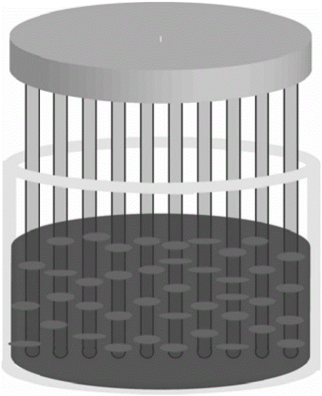
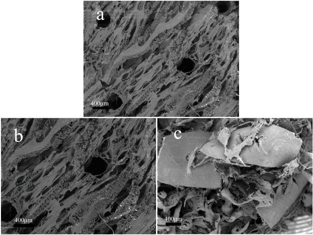
[0094] Use a 500 μm diameter acupuncture needle to place the cell amniotic membrane in step (1) in accordance with figure 1 The model shown (diameter 1.5 cm) is arranged (5 x 8...
PUM
| Property | Measurement | Unit |
|---|---|---|
| Area | aaaaa | aaaaa |
| Diameter | aaaaa | aaaaa |
| Diameter | aaaaa | aaaaa |
Abstract
Description
Claims
Application Information
 - R&D
- Intellectual Property
- Life Sciences
- Materials
- Tech Scout
- Unparalleled Data Quality
- Higher Quality Content
- 60% Fewer Hallucinations
Browse by: Latest US Patents, China's latest patents, Technical Efficacy Thesaurus, Application Domain, Technology Topic, Popular Technical Reports.
© 2025 PatSnap. All rights reserved.Legal|Privacy policy|Modern Slavery Act Transparency Statement|Sitemap|About US| Contact US: help@patsnap.com