Device and method for achieving preoperative assessment of congenital heart disease operation plan based on 3D printing
A 3D printing and surgical plan technology, applied in the field of medical image processing, can solve the problems of increasing the mental and economic burden of patients and their families, high medical expenses, increasing the suffering of patients, etc., to facilitate direct observation, improve the possibility of patient survival, Avoid painful effects
- Summary
- Abstract
- Description
- Claims
- Application Information
AI Technical Summary
Problems solved by technology
Method used
Image

Examples
Embodiment 1
[0036] At present, in clinical practice, in order to obtain the physiological parameter data of the heart and collateral vessels of patients with congenital heart disease, doctors use invasive interventional methods to obtain relevant parameters of congenital heart disease in newborns. Models, let alone simulate surgical operations.
[0037] With the development and innovation of computer science and technology, the computer-dependent medical image processing technology has been developed and improved, and the heart segmentation technology based on CT data has been greatly improved. The heart model obtained by using the active contour model algorithm to help The doctor intuitively understands the patient's condition.
[0038]Aiming at the existing technology, the present invention has carried out research and development, using 3D technology and medical image segmentation technology to provide a device for preoperative evaluation of congenital heart disease operation plan by m...
Embodiment 2
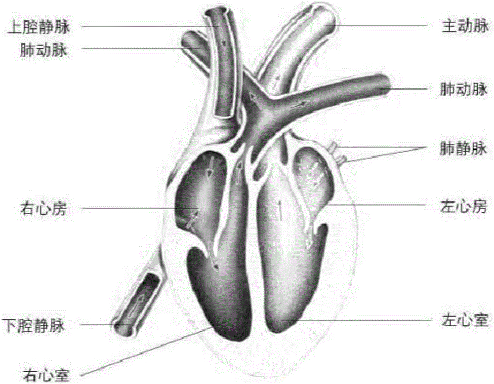
[0046] The overall structure of the device for preoperative evaluation of the congenital heart disease operation plan by means of 3D printing is the same as in Example 1. In the present invention, the three-dimensional structural model of the heart and collateral blood vessels of a normal person or patient is printed and connected with the corresponding blood vessel positions of the extracorporeal circulation machine. They are respectively connected with extracorporeal circulation machines through blood vessel branches. The 3D-printed three-dimensional structure model is connected to the extracorporeal circulation machine, and the connection nodes refer to Figure 6 , respectively, the inferior vena cava is 3 mm from the outlet of the right atrium, at a, the superior vena cava is 3 mm from the outlet of the right atrium, at b, the right pulmonary artery is 3 mm from the outlet of the right ventricle, at c, the aorta is 3 mm from the outlet of the left ventricle, and at d; The ...
Embodiment 3
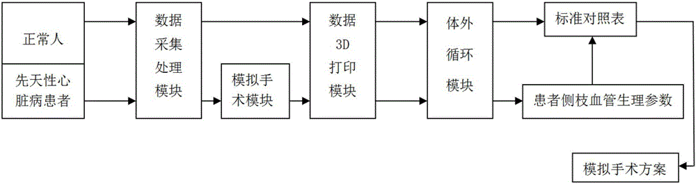
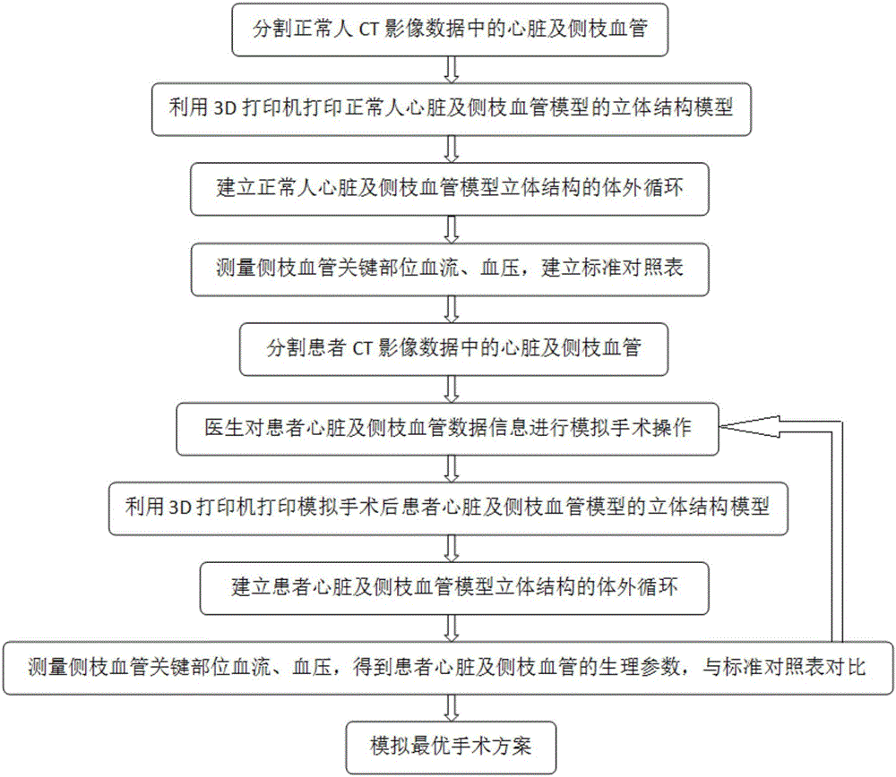
[0048] The overall structure of the device for preoperative evaluation of the congenital heart disease operation plan by means of 3D printing is the same as that of Embodiment 1-2. In the present invention, the data 3D printing module first prints out The three-dimensional structure of the normal human heart and collateral vessels. Connect the three-dimensional structure of the normal human heart and collateral vessels to the extracorporeal circulation machine to form an extracorporeal circulation module. The extracorporeal circulation module obtains the physiological parameters of the key parts of the normal human heart and collateral vessels, and establishes a standard comparison table , the standard comparison table is first stored in the physiological parameters of normal people.
[0049] Then the patient's heart and collateral blood vessel data pass through the data acquisition and processing module to obtain the segmented patient's heart and collateral blood vessel data i...
PUM
 Abstract
Description
Claims
Application Information
 - R&D
- Intellectual Property
- Life Sciences
- Materials
- Tech Scout
- Unparalleled Data Quality
- Higher Quality Content
- 60% Fewer Hallucinations
Browse by: Latest US Patents, China's latest patents, Technical Efficacy Thesaurus, Application Domain, Technology Topic, Popular Technical Reports.
© 2025 PatSnap. All rights reserved.Legal|Privacy policy|Modern Slavery Act Transparency Statement|Sitemap|About US| Contact US: help@patsnap.com