Anti-dermatophyte specific yolk antibody, preparation method and application thereof

A dermatophyte and egg yolk antibody technology, applied in skin care preparations, microorganism-based methods, biochemical equipment and methods, etc., can solve problems such as low specificity, achieve no toxic side effects, easy mass production, Strong specific effect

Inactive Publication Date: 2017-02-15
GUANGDONG UNIV OF TECH
View PDF4 Cites 6 Cited by
  • Summary
  • Abstract
  • Description
  • Claims
  • Application Information

AI Technical Summary

Problems solved by technology

Both of these two bacteria belong to the genus Trichophyton, and the fungal structures are similar, so the serum antibodies obtained from the two bacteria have cross-reactivity, and the specificity is not strong

Method used


Image

  • Anti-dermatophyte specific yolk antibody, preparation method and application thereof
  • Anti-dermatophyte specific yolk antibody, preparation method and application thereof
  • Anti-dermatophyte specific yolk antibody, preparation method and application thereof

Examples


Embodiment 1

[0024] Example 1: Preparation of dermatophyte protein antigen

[0025] (1) Cultivate Trichophyton rubrum and Trichophyton mentagrophytes by SDA solid medium, then inoculate in liquid culture medium and culture in large quantities, centrifuge, wash with PBS, obtain a large amount of hyphae;

[0026] (2) Extraction of cell wall protein: For (1) cultured mycelium, use liquid nitrogen to grind, add PBS to dissolve, add 10mM EDTA, 10mM PMSF to inhibit protease; , centrifuged to obtain the cell wall; add 4°C, 0.1N NaOH to the cell wall precipitate, stir at 4°C for 24h, 4°C, 12000g, and centrifuge for 10min to obtain the filtrate, neutralize the filtrate with pre-cooled 1mol / l HCl; The filtrate was dialyzed with 50mM Tris-Hcl for three days to obtain the dermatophyte cell wall protein antigen. Dilute the dermatophyte cell wall protein antigen with PBS buffer solution to a 1 mg / ml dermatophyte cell wall protein antigen solution for later use.

Embodiment 2

[0027] Embodiment 2: the preparation of anti-dermatophyte egg yolk antibody

[0028] (1) Immunization of laying hens with anti-dermatophyte antigen

[0029] Select 12 laying hens at the age of 20 weeks; the dermatophyte cell wall protein antigen solution obtained in Example 1 and Freund's complete adjuvant are mixed and emulsified according to the volume ratio of 1:1, and the immunization method of subcutaneous multi-point injection is used to treat each hen. The chicken was injected with 0.5ml; two weeks after the first immunization, the first booster immunization was carried out, and an equal volume of Freund's incomplete adjuvant was mixed and emulsified with the dermatophyte cell wall protein antigen solution, and each hen was immunized by subcutaneous multi-point injection. The chickens were injected with 0.5ml, and booster immunization was carried out every 15 days. A total of 4 booster immunizations were performed. The immunized eggs were collected from the first booste...

Embodiment 3

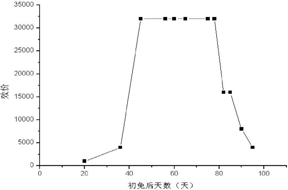
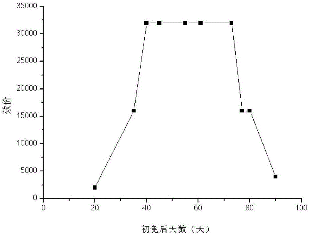
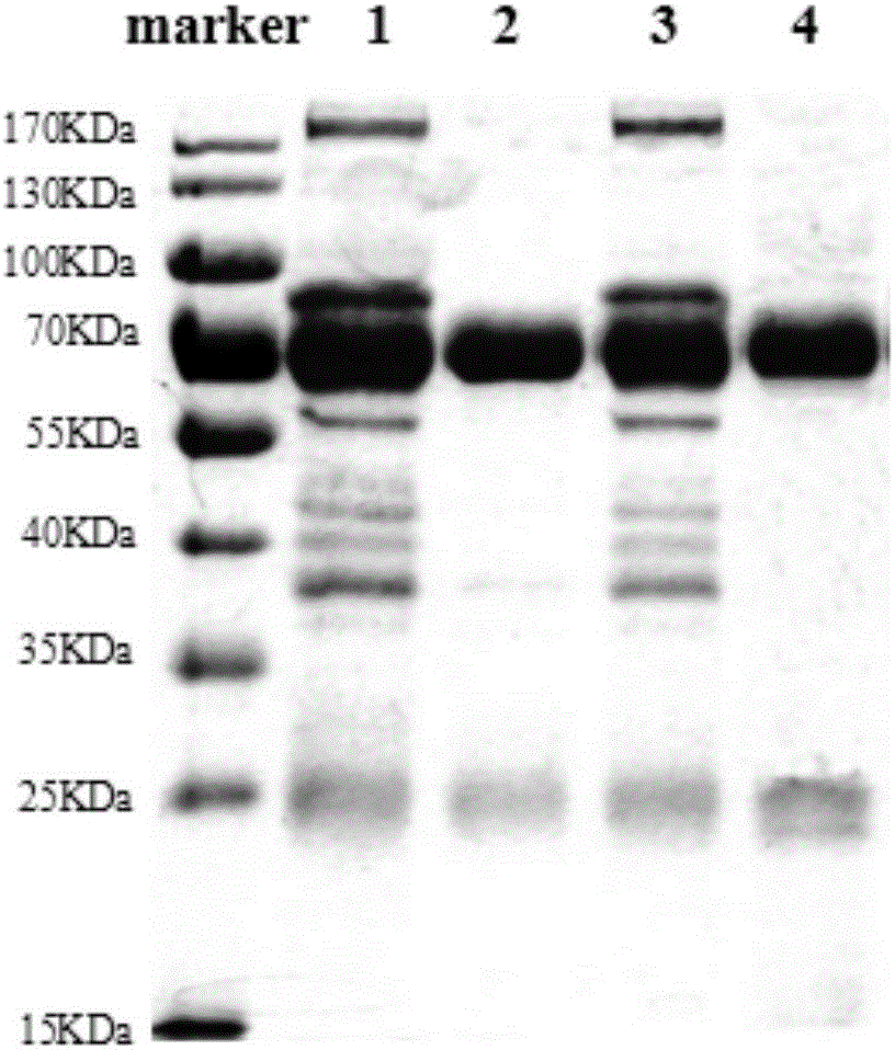
[0033] Embodiment 3: Titer detection of anti-dermatophyte specific egg yolk antibody

[0034]ELISA was used to detect the titers of egg yolk antibodies obtained by immunization with various antigens. Use a 96-well enzyme-labeled plate, add 100 μl of whole bacterial antigens coated with Trichophyton rubrum and Trichophyton mentagrophytes to each well of two enzyme-labeled plates, cover with a sealing film, incubate at 37°C for 2 hours, and then shake dry , washed 3 times with PBST, patted dry with absorbent paper; added 100 μl / well 30 mg / ml skimmed milk powder to block, incubated at 37°C for 1 hour, then dried, washed 3 times with PBST, patted dry with absorbent paper; The IgY obtained from the immunization of two cell wall proteins of Trichophyton mentagrophytes and Trichophyton mentagrophytes was serially diluted, and 100 μl / well was added to the microtiter plate, and non-immunization was added to the last line as a blank control, incubated at 37°C for 1 hour, then shaken dry...


PUM


Abstract

The invention belongs to the field of biotechnology, and discloses an anti-dermatophyte specific yolk antibody, a preparation method and the application thereof. The preparation method comprises the following steps: 1) selecting two major pathogenic bacteria of tinea, and culturing the two major pathogenic bacteria to respectively extract the bacteria cell wall-associated proteins of the bacteria; 2) preparing antigen-immunized hens based on obtained proteins for five times in total so as to obtain eggs, extracting and purifying egg yolk antibodies from the eggs; 3) preparing an anti-dermatophyte formulation based on obtained IgY. According to the technical scheme of the invention, by utilizing two cell wall-associated proteins of tinea pathogenic bacteria, the prepared anti-dermatophyte egg yolk antibody is high in specificity and free of toxic and side effects, which can effectively inhibit dermatophyte. Meanwhile, the preparation method is easy for mass production, low in cost, and simple in process. based on the method, the obtained formulation is very safe and can prevent dermatophyte diseases.

Description

technical field [0001] The invention belongs to biopharmaceutical technology, and in particular relates to a specific yolk antibody against dermatophytes and its preparation method and application. Background technique [0002] Fungi are widespread throughout the world, mainly in soil and on humans and animals. Wherein, the common disease that is infected by fungus is ringworm of the skin. For example: athlete's foot, psoriasis, etc. During the process of proliferation, dermatophytes penetrate deep into the dermis, stimulating the nervous system of the dermis and causing itching symptoms; local tissue fluid gathers, and the skin tissue is easily damaged by secondary bacterial infections, leading to inflammatory reactions. A series of skin histopathological phenomena caused are called dermatophytosis. Dermatophytosis is a common disease in humans, and at least 10-20% of the world's population may be infected with these pathogens. In recent years, due to the wide applicati...

Claims


Application Information

Patent Timeline
Patent Type & Authority Applications(China)
IPC IPC(8): C07K16/14C07K16/02A61K39/39A61K9/06A61K9/12A61P31/10A61K8/64A61Q19/10A61Q17/00C12R1/645
CPCA61K8/64A61K9/06A61K9/12A61K2039/505A61Q17/005A61Q19/10C07K16/02C07K16/14C07K2317/73
Inventor 赵肃清胡青青焦洛莹夏娜娜吕瑞梁雨欣
Owner GUANGDONG UNIV OF TECH
Why Eureka
Try Eureka