
Body fluid circulation DNA quantitative determination method and kit

A quantitative detection method and real-time fluorescence quantitative technology, applied in biochemical equipment and methods, microbial determination/inspection, etc., can solve the problems affecting the precision and stability of quantitative results, low detection sensitivity, and large differences between tubes.

Active Publication Date: 2017-02-15
JIANGSU CODE BIOMEDICAL TECH CO LTD
View PDF3 Cites 4 Cited by
  • Summary
  • Abstract
  • Description
  • Claims
  • Application Information

AI Technical Summary

Problems solved by technology

Fluorescent quantitative PCR external standard method is a single-plex fluorescent quantitative PCR for human housekeeping genes. A standard curve is established through an external standard of known concentration, and quantitative analysis is performed on the sample to be tested. Since the standard and the sample to be tested are in different test tubes The operation is carried out in the medium, and the difference between the tubes is large, which affects the precision and stability of the quantitative results.
The PicoGreen fluorescence method uses the PicoGreen fluorescent dye to bind non-specifically to double-stranded DNA. After the combination, the fluorescence intensity increases significantly. A standard curve is established through an external standard with a known concentration, so as to perform quantitative analysis on the sample to be tested. Because it is a direct detection without DNA Amplification, the detection sensitivity of this method is lower than that of fluorescent quantitative PCR method, and there are large differences between tubes, which affect the precision and stability of quantitative results
In order to solve the above problems, the inventor designed the plasma circulating DNA internal standard method for quantitative detection in the early stage, see CN1811387 for details, although it is improved to a certain extent compared with the PCR external standard method and PicoGreen fluorescence method, but due to the problems of internal standard sequence and primer sequence design , causing the length of the target fragment to be amplified by the internal standard and the target gene to be different, resulting in inconsistent amplification efficiencies of the two in the dual fluorescent PCR reaction process, which still has a significant impact on the quantitative detection results

Method used


Image

  • Body fluid circulation DNA quantitative determination method and kit
  • Body fluid circulation DNA quantitative determination method and kit
  • Body fluid circulation DNA quantitative determination method and kit

Examples


Embodiment 1

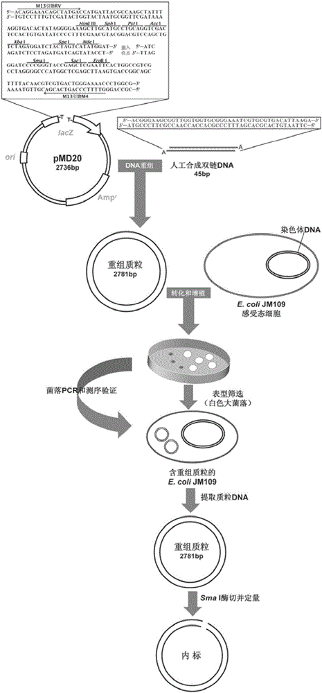
[0036] (1) Synthesis of internal standard

[0037] Artificially synthesized linear double-stranded DNA fragments with "A" cohesive ends, the sequence is as follows:

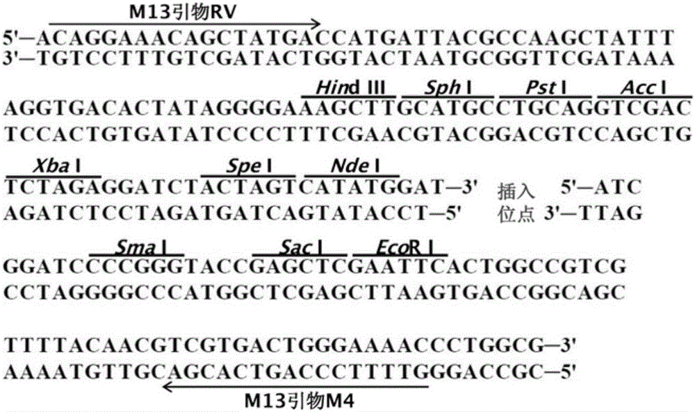
[0038] 5'-ACGGGAAGCGGTTGGTGGTGCGGGAAATCGTGCGTGACATTAAGA-3'

[0039] 3’-ATGCCCTT CGCCAACCACCACGCCC TTT AGCACGCACTGTAATTC-5’

[0040] Dilute the two artificially designed complementary oligonucleotides to 20 μmol / L with Tris-EDTA buffer, mix them with equal volumes, put them in a boiling water bath at 100°C for 10 minutes, and then let them cool naturally at room temperature to obtain linear double-stranded DNA fragments; Recombine the linear double-stranded DNA fragment into the plasmid vector T-Vector pMD20 (for the insertion site, see figure 2 ), transfected into E.coli JM109 competent cells, cultured in vitro and inoculated on L-agar plate medium containing X-Gal, IPTG, Amp, directly screened large white colonies containing recombinant clones, and then sequenced by M13 Universal primers RV and M4 were used ...

Embodiment 3

[0077] Example 3 Detection of Plasma Samples from Healthy Examiners

[0078] 400 healthy subjects, aged 25-60 years, with an average age of 43 years, 200 males and 200 females. Collect EDTA-K 2 Anticoagulated peripheral venous blood was separated by two-step centrifugation to obtain cell-free plasma samples. The plasma DNA was extracted using the magnetic bead method kit, and quantitatively determined by the method of the present invention, the internal standard method in the early stage, the traditional external standard method and the fluorescence method. The results Such as Figure 6 . The detection result of the method of the present invention is significantly higher than that of the previous internal standard method, traditional external standard method and fluorescence method, and the difference is statistically significant (P<0.001). The present invention eliminates the measurement error caused by the loss of nucleic acid extraction through the internal reference fun...

Embodiment 4

[0079] Example 4 Detection of healthy person's urine sample

[0080] 1000 healthy persons, aged 25-60 years, with an average age of 42 years, 500 males and 500 females. Collect morning urine specimens, two-step centrifugal separation to obtain cell-free urine specimens, use the magnetic bead method kit to extract urine DNA, and use the method of the present invention for quantitative determination. The results are as follows: Figure 7 . The quantitative range of urine DNA in healthy individuals ranged from 1.0 to 296.4 ng / ml (median: 18.1 ng / ml; interquartile range: 30.5 ng / ml), of which 84% had urine DNA below 50 ng / ml, and 12 % was between 50-100ng / ml, 4% exceeded 100ng / ml, and the urine DNA level was positively skewed in the 1000 cases of healthy people ( Figure 7 ). There was no statistically significant difference in urine DNA content between different sex or age groups.

[0081] Nanjing Meclelin Biomedical Technology Co., Ltd.

[0082] Method and Kit for Quantit...


PUM


Abstract

The invention discloses a body fluid circulation DNA quantitative determination method and kit. The method includes steps of selecting artificial single copy house-keeping gene Beta-action as the detection target gene, and artificially compounding a dual-chain DNA segment with length of 45 bp, wherein it includes sequences with the same 25 bp and Beta-action genes, and recombining to a plasma carrier T-Vector pMD 20 and obtaining the recombined plasmid DNA as an interior label, and feeding a certain quantity of interior label to a body fluid sample to be tested; synchronously storing with the body fluid circular DNA, and performing nucleic acid extraction and purification, and fluorogenic quantitative PCR amplification, so as to perform the body fluid circulation DNA quantitative analysis. Through setting up a special internal label, the method eliminates the detection bias caused by nucleic acid degradation, extraction loss and PCR amplification efficiency before and in body fluid sample analysis, and can perform more stable and exact quantitative determination on the body fluid circulation DNA.

Description

technical field [0001] The invention belongs to the field of biological sample detection, and relates to a method for quantitatively detecting body fluid circulating DNA and a kit. Background technique [0002] Circulating DNA, also known as cell-free DNA, exists in body fluids, such as extracellular DNA in blood (plasma or serum), urine, cerebrospinal fluid, and synovial fluid. Quantitative detection results of circulating DNA in body fluids are affected by many factors, which will cause large deviations, among which the loss and degradation of DNA during nucleic acid extraction and long-term storage are the two most important influencing factors. [0003] Due to the low content of circulating DNA in body fluid samples, quantitative analysis using gene amplification methods is required. The detection of gene amplification requires the extraction of nucleic acid. Due to the loss of nucleic acid during the extraction process, the quantitative results cannot reflect the true ...

Claims


Application Information

Patent Timeline
IPC IPC(8): C12Q1/68
CPCC12Q1/6851C12Q2561/113C12Q2561/101C12Q2537/143C12Q2545/107
Inventor 潘世扬陈丹徐建黄珮珺王芳
Owner JIANGSU CODE BIOMEDICAL TECH CO LTD
Learn More