Chitosan derivative with endosome escape function as well as preparation method and application thereof

A technology of chitosan derivatives and escape function, which can be applied to medical preparations with non-active ingredients, medical preparations containing active ingredients, and pharmaceutical formulas, etc., and can solve the problems of poor drug loading effect and poor effect

Active Publication Date: 2017-05-10
ZHEJIANG PHARMA COLLEGE
View PDF2 Cites 0 Cited by
  • Summary
  • Abstract
  • Description
  • Claims
  • Application Information

AI Technical Summary

Problems solved by technology

[0006] At present, there are many researches on compounds with endosome escape function, but there are still some problems, such as poor effect, poor drug loading effect, etc.

Method used


Image

  • Chitosan derivative with endosome escape function as well as preparation method and application thereof
  • Chitosan derivative with endosome escape function as well as preparation method and application thereof
  • Chitosan derivative with endosome escape function as well as preparation method and application thereof

Examples


Embodiment 1

[0027] Embodiment 1 material synthesis

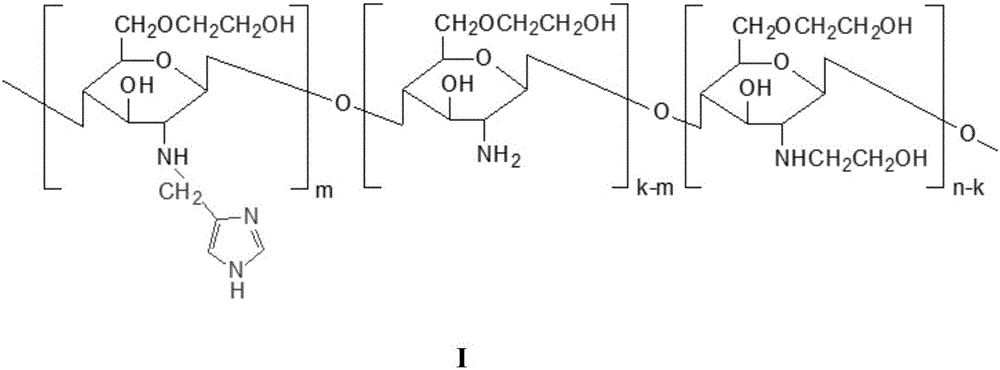
[0028] The material characterization method is as follows: by 1 H NMR and organic element analysis were used to determine the degree of substitution of imidazolylmethyl and hydroxyethyl, respectively. Determination of Critical Micelle Concentration of MHC Using Pyrene Fluorescence Spectrometry

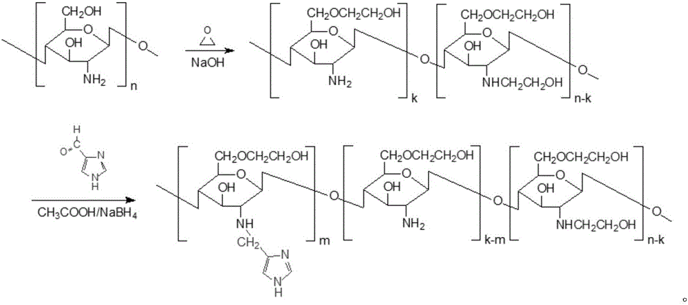
[0029] 1. Synthesis of hydroxyethyl chitosan (HE-Cs): Weigh 1 gram of chitosan (50KDa 100KDa), add 10ml of 2% HAc, stir until completely dissolved, then add 10ml of 50% NaOH. Afterwards, the temperature was raised to 40° C. for 12 hours, and 10 ml of ethylene oxide was added after cooling down with ice, and the temperature was raised to 50° C. for 18 hours. Cool to room temperature and add 5mol / L HCl to adjust the pH to neutral. The obtained product was centrifuged at 3000 rpm for 10 min, filtered through a 0.8 μm microporous membrane, dialyzed and freeze-dried to obtain the product.

[0030] Elemental analysis shows that different ethylene ...

Embodiment 2

[0038] Embodiment 2 drug loading process optimization

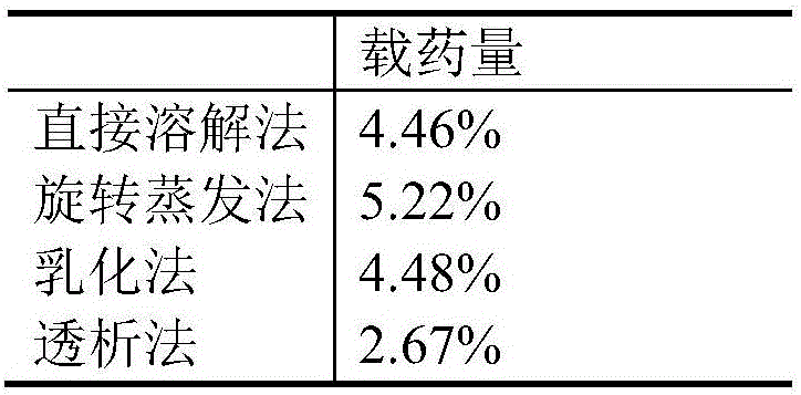
[0039] Taking quercetin as a model drug, the micelles prepared by direct dissolution method, rotary evaporation method, dialysis method and emulsification method with different drug loading were compared. Different solvents (ethanol, dimethyl sulfoxide, methanol), carrier concentrations (1%, 0.67%, 0.5%), and drug-carrier ratios (1:25, 1:10) of quercetin were compared using a single factor method. , 1:5) on the drug loading, the process was optimized with the drug loading as an index, and the drug-loading micelles reaching a certain concentration were made. The results are shown in Table 3 and Table 4 below.

[0040] Table 3 Drug-loaded micelles obtained by different preparation methods

[0041]

[0042] Table 4 single factor inspection process optimization results

[0043]

Embodiment 3

[0044] Example 3 Cellular uptake and intracellular endosome escape

[0045] Fluorescent probes with coumarin-6 (C6) as hydrophobic drugs are entrapped in micelles, and are observed and evaluated by laser confocal.

[0046] For the convenience of microscopic observation, MDA-MB-231 cells were cultured in DMEM for 24 hours in a 24-well plate. After incubation with micelles for 4 hours, the cells were washed three times with PBS and fixed with 4% paraformaldehyde. Cell nuclei were stained with Hoechst 33258 before fluorescence photography, and the cellular uptake of micelles was observed by fluorescence microscopy.

[0047] CLSM was used to observe the subsequent internalization of micelles and endosomal escape. MDA-MB-231 cells were cultured in a glass-bottomed culture dish for 24 hours, washed three times with PBS at 2 hours, 4 hours and 12 hours after adding C6-MHC micelles, and then used LysoTracker TM Red staining, 4% paraformaldehyde fixation, CLSM observation.

[0048]...


PUM


Abstract

The invention discloses a chitosan derivative with an endosome escape function as well as a preparation method and application thereof. The chemical structure of the derivative is shown by a formula I in the specification, wherein n is the polymerization degree of chitosan, m / n is the methyl substitution degree of imidazole, and k / n is the proportion of amino on the unit saccharide ring not substituted by hydroxyethyl. Compared with the prior art, an amphiphilic chitosan derivative is prepared by a simple synthesis way, and endosome escape is realized through a proton sponge effect. The obtained carrier material has certain drug loading ability and can help a drug realize the endosome escape function.

Description

technical field [0001] The invention relates to a chitosan derivative with endosome escape function, a preparation method and application thereof, and belongs to the technical field of biomedical materials. Background technique [0002] Cancer has become a frequently-occurring and common disease that seriously threatens human life and health worldwide. The current clinical treatment of cancer includes drug therapy, radiotherapy and surgery, among which chemotherapy plays a dominant role in cancer treatment. However, due to the low selectivity of chemical drugs, when they kill tumor cells and inhibit the growth and reproduction of tumor cells, they also have the same effect on normal tissues, organs and cells, resulting in serious adverse reactions and even failure of chemotherapy. Although the nano drug delivery system studied in recent years has tumor tissue targeting and can enter cells through the pinocytosis pathway, it will inevitably be phagocytized into endosomes, an...

Claims


Application Information

Patent Timeline
IPC IPC(8): C08B37/08A61K31/352A61K31/37A61K9/107A61K47/36
CPCA61K9/1075A61K31/352A61K31/37A61K47/36C08B37/003
Inventor 夏晓静胡英徐蓓华
Owner ZHEJIANG PHARMA COLLEGE
Why Eureka
Try Eureka