Urine exfoliated tumor cell micro-fluidic chip detection technology aiming at urothelium carcinoma

A microfluidic chip, cell technology, applied in the direction of tumor/cancer cells, animal cells, vertebrate cells, etc., can solve the problems of high work intensity, disordered cell composition, inconsistent cell size, etc., to improve detection accuracy, get rid of Effects of Subjective Experience Dependence

Pending Publication Date: 2017-06-20
ZHEJIANG UNIV +1
View PDF5 Cites 13 Cited by
  • Summary
  • Abstract
  • Description
  • Claims
  • Application Information

AI Technical Summary

Problems solved by technology

[0005] (3) The dyeing process is cumbersome, with high manual participation and high work intensity
[0008] (1) It is an invasive examination, and the patient suffers a lot;
[0009] (2) The tumor can only be observed when it develops to the naked eye, which is not conducive to early diagnosis.
[0013] (2) The pore diameter of the filter membrane is single, 7.5um; however, the cell composition in the urine of patients with urothelial carcinoma is complex, in addition to tumor cells, there are also normal exfoliated urothelial cells, white blood cells, red blood cells, etc., these The cell sizes are inconsistent. If a single pore size is used, the cell components in the field of view will still be confused, which will interfere with subsequent judg

Method used


Image

  • Urine exfoliated tumor cell micro-fluidic chip detection technology aiming at urothelium carcinoma
  • Urine exfoliated tumor cell micro-fluidic chip detection technology aiming at urothelium carcinoma
  • Urine exfoliated tumor cell micro-fluidic chip detection technology aiming at urothelium carcinoma

Examples


preparation example Construction

[0075] 2. Preparation method of microfluidic chip

[0076] The microfluidic chip of the present invention can be prepared according to the standard process of polydimethylsiloxane (PDMS), using a conventional glass slide as a substrate, and bonding through plasma treatment.

[0077] PDMS is the English abbreviation of polydimethylsiloxane. Polydimethylsiloxane is a curable polymer. After the curable polymer is mixed with a curing agent, it can be cured and hardened after a period of time to obtain a microfluidic chip with a certain structure.

[0078] 3. The use of microfluidic chips

[0079] The microfluidic chip of the present invention can be used to capture or recover urine exfoliated cells.

[0080] 4. Method for capturing or recovering urine exfoliated cells

[0081] The method of capturing or recovering urine exfoliated cells of the present invention includes steps: (1) capturing: the urine sample to be processed or the suspension of urine sediment enters the microfl...

Embodiment 1

[0112] Example 1 Microfluidic chip captures urine exfoliated cells

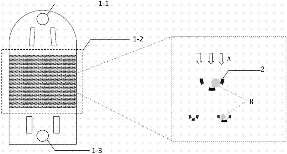
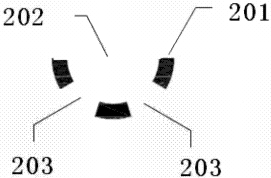
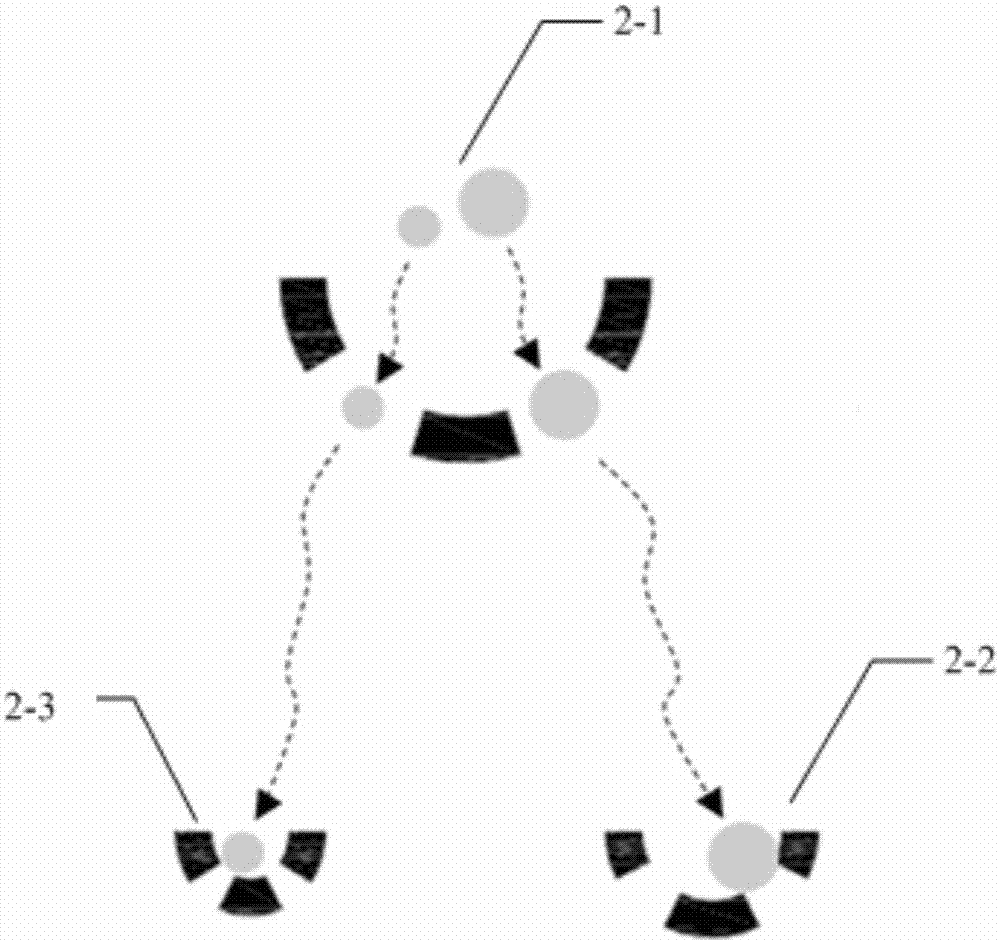
[0113] Such as Figure 1~5 As shown, the microfluidic chip of the present invention includes a sample inlet 1-1, a cell capture area 1-2 and a sample outlet 1-3 connected in sequence along the liquid flow direction A. The cell capture area 1-2 is provided with a plurality of cell sorters 2, such as figure 2 As shown, the cell sorter 2 is composed of three columnar projections 201 arranged in an arc as a whole. There are gaps between the columnar projections. The arc-shaped openings 202 serve as liquid inlets. The gap 203 serves as a liquid outlet, and the two liquid outlets are symmetrically distributed. The cell capture area 1-2 is equipped with cell sorters 2 of three different sizes, namely the first size cell sorter 2-1, the second size cell sorter 2-2 and the third size cell sorter 2-3, the size of the cell sorter 2-1 of the first size, the cell sorter 2-2 of the second size and the cell sorter 2-3 o...

Embodiment 2

[0118] Example 2 Determining Urinary Exfoliation Tumor Cells by Immunofluorescence

[0119] After the urine exfoliated cells are captured by the microfluidic chip in Example 1, as Image 6 As shown, the cells of different sizes are regularly distributed inside the chip, the field of view is clear, and the cell shape is intact, which is especially suitable for the next analysis.

[0120] In this example, immunofluorescence method was used to distinguish urinary exfoliated tumor cells from normal urothelial cells.

[0121] Immunofluorescence staining step: Paraformaldehyde passes through the microfluidic chip with urine exfoliated cells at a flow rate of 0.5-4ml / h for 30 minutes; PBS passes through the microfluidic chip at a flow rate of 0.5-4ml / h for 3 minutes. -5min; the Triton X-100 solution with a concentration of 0.1% passes through the microfluidic chip at a flow rate of 0.5 to 4ml / h for 10 minutes; PBS passes through the microfluidic chip at a flow rate of 0.5 to 4ml / h a...


PUM


Abstract

The invention belongs to the field of biological fluid detection in vitro, and concretely relates to a urine exfoliated tumor cell micro-fluidic chip detection technology aiming at urothelium carcinoma. The micro-fluidic chip comprises a sample inlet, a cell capturing area and a sample outlet which are connected sequentially, wherein the cell capturing area is provided with a plurality of cell sorting devices; each cell sorting device is formed by three columnar bulges arranged in an arc shape; a gap exists between every two columnar bulges; an arc-shaped opening is used as a liquid flow inlet; gaps at two sides of each middle columnar bulge are used as liquid flow outlets which are symmetrically distributed. According to the urine exfoliated tumor cell micro-fluidic chip detection technology provided by the invention, various cells in urine are separated and captured, multiple downstream cytology staining methods can be compatible for realizing cell recognition, the detection accuracy of urine exfoliated tumor cells in human urine is improved, the dependency on subjective experiences of pathology doctors in a conventional method is avoided, and the whole process is noninvasive. In addition, a lossless cell capturing and recovering manner lays a foundation on downstream molecular biology analysis.

Description

technical field [0001] The invention belongs to the field of extracorporeal detection of biological bodies, and in particular relates to a microfluidic chip detection technology for urinary exfoliation tumor cells for urothelial cancer. Background technique [0002] 1. Active clinical technology-urinary exfoliation cytology examination: Urinary exfoliation cytology examination (Cytology) is currently a commonly used clinical method for the detection of urothelial carcinoma. In order to find tumor cells, this method relies on the subjective observation of pathologists on smears (judging cells based on factors such as cell nuclei, chromatin, and cytoplasm), which mainly has the following disadvantages: [0003] (1) The detection sensitivity is low, generally only 30-50%. [0004] (2) The film reading depends entirely on the subjective experience of pathologists, and the judgment results of different pathologists may be inconsistent. [0005] (3) The dyeing process is cumbers...

Claims


Application Information

Patent Timeline
IPC IPC(8): C12M1/00C12N5/09C12Q1/02
CPCG01N33/5044G01N33/5091C12N5/0693
Inventor 金百冶韩平畴傅广候陈安琪
Owner ZHEJIANG UNIV
Why Eureka
Try Eureka