
Identification method of EB virus infected lymphocyte subpopulation and application thereof

A technology of lymphocytes and identification methods, applied in the field of identification of EB virus-infected lymphocyte subsets, can solve the problems of poor detection of EB virus

Active Publication Date: 2017-07-14
BEIJING FRIENDSHIP HOSPITAL CAPITAL MEDICAL UNIV
View PDF3 Cites 10 Cited by
  • Summary
  • Abstract
  • Description
  • Claims
  • Application Information

AI Technical Summary

Problems solved by technology

[0005] The first object of the present invention is to provide a kind of identification method of Epstein-Barr virus-infected lymphocyte subsets, and the second object of the present invention is to provide the application of the identification method of EB virus-infected lymphocyte subsets, to alleviate the existing technology. Existing technical problems of poor detection of EB virus

Method used


Image

  • Identification method of EB virus infected lymphocyte subpopulation and application thereof
  • Identification method of EB virus infected lymphocyte subpopulation and application thereof
  • Identification method of EB virus infected lymphocyte subpopulation and application thereof

Examples


Embodiment 1

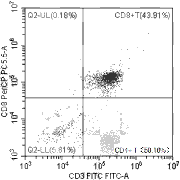
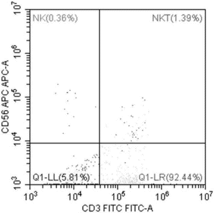
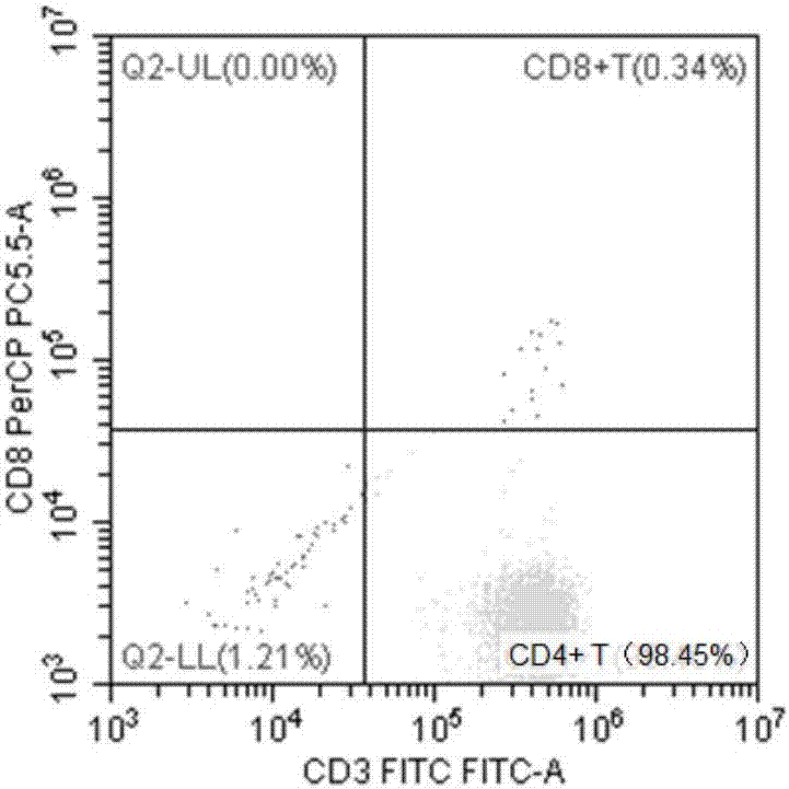
[0045] This embodiment provides a method for identifying a subset of lymphocytes infected with Epstein-Barr virus based on magnetic beads, comprising the following steps:

[0046] 1. Extraction of human peripheral blood mononuclear cells (HPBMC): 6 mL of fasting venous blood was placed in a sterile test tube anticoagulated with EDTA, diluted 1-fold with PBS, and then separated from PBMC with lymphocyte separation medium (2000 rpm for 20 minutes). Prepare the buffer solution (MACS BSA Stock Solution (#130-091-376) and autoMACS Rinsing Solution (#130-091-222) in a ratio of 1:20) during centrifugation, and put it in a 4-degree refrigerator to cool down;

[0047] 2. Absorb the middle buffy coat layer, add 10mL PBS, 1400rpm, 10min to wash and precipitate PBMC;

[0048] 3. Discard the supernatant, add 2mL erythrocyte lysate (Solebo erythrocyte lysate product number: Cat#R1010) to lyse, mix well, and incubate at room temperature for 5 minutes;

[0049] 4. After fully mixing, absorb ...

Embodiment 2

[0080] The present embodiment provides the identification method of the Epstein-Barr virus-infected lymphocyte subsets based on flow cytometry, comprising the following steps:

[0081] 1. Take 6 mL of peripheral blood, anticoagulate with heparin, dilute to 12 mL with PBS, and mix well;

[0082] 2. Slowly add the diluted blood above the liquid surface of 15mL lymphocyte separation solution along the wall of the test tube, do not use too much force, so as not to cause the blood to mix with the separation solution and maintain a clear layered state;

[0083] 3. Centrifuge at 2000rpm for 20min at 18-20°C. After centrifugation, it can be seen that the blood in the test tube is clearly divided into 4 layers, the upper layer is the plasma layer, and the middle layer is the separation liquid layer (the mononuclear cells are in the middle of the plasma layer and the separation liquid layer) , the bottom layer is the erythrocyte layer, and the granulocyte layer is above the erythrocyte ...


PUM


Abstract

The invention provides an identification method of an EB virus infected lymphocyte subpopulation and an application thereof and relates to the technical field of medical detection. The identification method of the EB virus infected lymphocyte subpopulation provided by the invention can accurately position the cell type of EBV infection by sorting monocytes of peripheral blood of an EBV infected patient in combination with a flow cytometry and a real-time fluorescent quantitative PCR technology, and is helpful for a clinician to formulate a therapeutic regimen for the cell type with EBV infection so as to efficiently and accurately treat diseases caused by EBV infection. In addition, by applying the identification method provided by the invention, target cells infected with EBV can be determined, preparation of related therapeutic drugs can be guided, and the targeted drugs are prepared with a clear target.

Description

technical field [0001] The invention relates to the technical field of medical detection, in particular to a method for identifying lymphocyte subsets infected with Epstein-Barr virus and its application. Background technique [0002] Epstein-Barr virus (EBV) belongs to the fourth type of human herpes virus and was discovered by Epstein and Barr in 1964 when they were studying malignant lymphoma in African children. Epstein-Barr virus is a lymphotropic virus, which is one of the most commonly infected viruses in humans, infecting more than 90% of the world's population. Epstein-Barr virus has been designated as a class I carcinogen by the International Cancer Research Center in 1997 because of its high infection rate and carcinogenic rate. [0003] At present, there are some detection kits for Epstein-Barr virus, but most of them detect Epstein-Barr virus infection in whole blood and are not targeted. [0004] In view of this, the present invention is proposed. Contents ...

Claims


Application Information

Patent Timeline
IPC IPC(8): C12Q1/70C12Q1/68G01N15/14G01N21/64C12R1/93
CPCC12Q1/705C12Q2600/136G01N15/14G01N21/6486
Inventor 王昭高卓王旖旎张嘉王晶石吴林
Owner BEIJING FRIENDSHIP HOSPITAL CAPITAL MEDICAL UNIV
Learn More