
Wide field super-resolution microscopic imaging method and wide field super-resolution microscopic imaging apparatus based on total internal reflection structure illumination

A technology of structured light illumination and microscopic imaging, which is applied in the direction of measuring devices, material analysis and material analysis through optical means, can solve the problems of limiting imaging speed, achieve improved imaging speed, high contrast of interference fringes, and easy operation Effect

Active Publication Date: 2017-07-14
ZHEJIANG UNIV
View PDF4 Cites 18 Cited by
  • Summary
  • Abstract
  • Description
  • Claims
  • Application Information

AI Technical Summary

Problems solved by technology

Although the requirements for the incident light power are low, the further improvement of the imaging speed is limited due to the raster scanning required for image processing

Method used


Image

  • Wide field super-resolution microscopic imaging method and wide field super-resolution microscopic imaging apparatus based on total internal reflection structure illumination
  • Wide field super-resolution microscopic imaging method and wide field super-resolution microscopic imaging apparatus based on total internal reflection structure illumination
  • Wide field super-resolution microscopic imaging method and wide field super-resolution microscopic imaging apparatus based on total internal reflection structure illumination

Examples


Embodiment 1

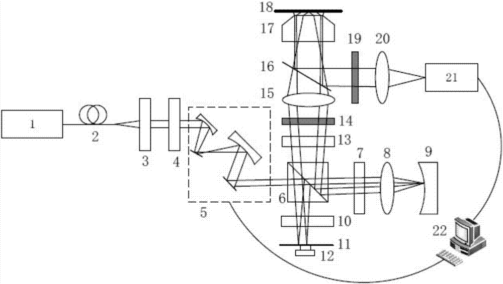
[0064] Such as figure 1 The shown wide-field super-resolution microscopy imaging device includes: laser 1, single-mode fiber 2, polarizer 3, half-wave plate 4, scanning galvanometer system 5, polarizing beam splitter 6, the first quarter A wave plate 7, a first convex lens 8, a concave mirror 9, a second quarter wave plate 10, a plane mirror 11, a piezoelectric ceramic 12, a third quarter wave plate 13, and a tangential polarizer 14. A second convex lens 15, a dichroic mirror 16, a microscope objective lens 17, a fluorescent sample to be measured 18, a filter 19, a third convex lens 20, a CMOS industrial camera 21, and a computer 22.

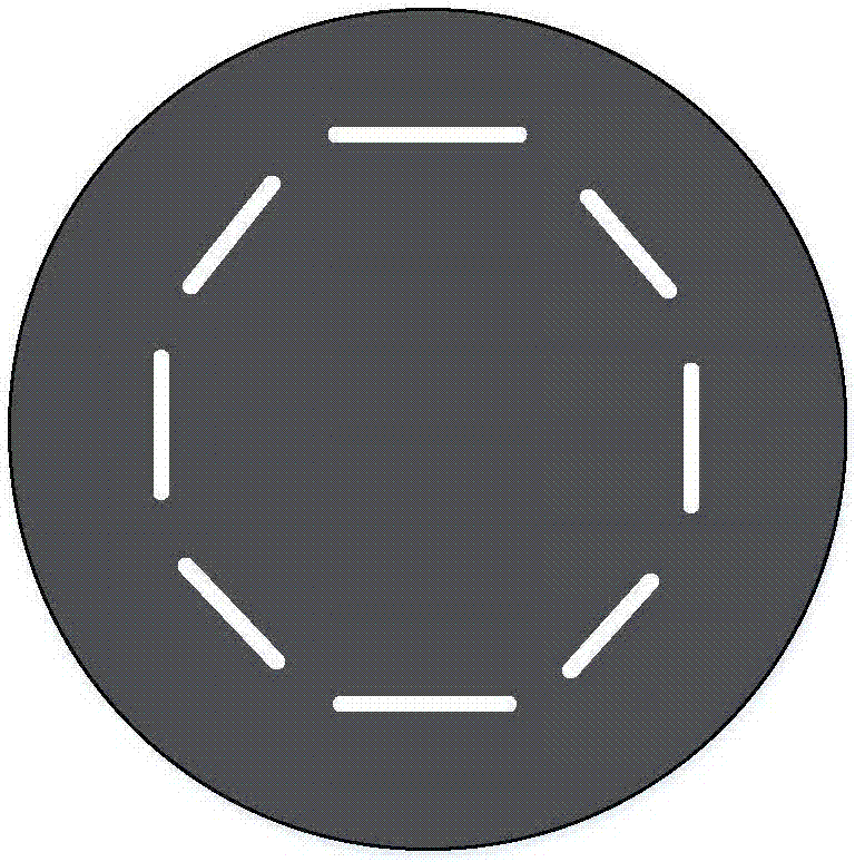
[0065] A laser 1 emits a laser beam, and a single-mode fiber 2 , a polarizer 3 and a half-wave plate 4 are sequentially placed on the optical axis of the optical path of the laser beam. The single-mode fiber 2 is used to filter the laser beam, the polarizer 3 is used to convert the outgoing laser light into linearly polarized light, and the hal...

Embodiment 2

[0076] Such as Figure 4 As shown, the wide-field super-resolution microscopic imaging device of this embodiment can also be realized by using a tangential light polarization converter. Figure 4 and figure 1 Compared, the polarized beam splitter 6 is replaced by the non-polarized beam splitter 23, and the first quarter wave plate 7, the second first quarter wave plate 10, the third first quarter wave plate are removed. A wave plate 13 and a tangential light polarizer 14 are added, and a tangential light polarization converter 24 is added at the same time. Since the tangential light polarization converter 24 requires the incident light to be point incident, it needs to be placed at the focal plane of the second convex lens 15 , that is, at the entrance pupil plane of the microscopic objective lens 17 . The non-polarizing beam splitter 23 splits the incident ray polarized light into two paths and enters the two interference arms of the off-axis Michelson interferometer withou...


PUM


Abstract

The present invention discloses a wide field super-resolution microscopic imaging method based on total internal reflection structure illumination. The wide field super-resolution microscopic imaging method comprises that a laser beam is divided into two linearly polarized light beams having symmetrical propagation directions and perpendicular vibration directions; the two linearly polarized light beams are converted into two tangential linearly polarized light beams, the two tangential linearly polarized light beams are projected on a fluorescent sample, total reflection is generated and mutual interference is generated to produce a fringe structure illumination pattern; the fluorescent signal emitted by the sample is collected to obtain fluorescent intensity information; by sequentially rotating the direction of the interference fringe of the structure illumination pattern, the phase of the interference fringe is changed a plurality of times in various directions to obtain a plurality of fluorescent intensity images under the corresponding phases in various directions; and data processing is performed on the plurality of the fluorescent intensity images, and re-construction is performed to obtain the super-resolution image. The present invention further discloses a wide field super-resolution microscopic imaging apparatus based on total internal reflection structure illumination. According to the present invention, the incident light energy utilization rate is high, the interference fringe contrast is high, and the resolution exceeding the diffraction limit under the low incident light power condition can be achieved.

Description

technical field [0001] The invention belongs to the field of optical super-resolution microscopic imaging, in particular to a wide-field super-resolution microscopic imaging method and device based on total internal reflection structured light illumination. Background technique [0002] Light microscopy plays an important role in life science research. However, due to the existence of the diffraction limit, that is, the ideal point object through the optical system forms a diffuse spot of finite size, resulting in the imaging resolution of ordinary optical microscopes being limited to about half the wavelength, that is, 200nm. [0003] In recent years, in order to break through the diffraction limit and observe the structure and function of cells at the nanoscale, researchers have proposed a variety of super-resolution optical microscopy techniques. For example, imaging methods based on single-molecule localization techniques, including light-activated localization microsco...

Claims


Application Information

Patent Timeline
Patent Type & Authority Applications(China)
IPC IPC(8): G01N21/64
CPCG01N21/6458
Inventor 匡翠方刘文杰陈友华朱大钊刘旭张克奇毛磊
Owner ZHEJIANG UNIV
Learn More