Multilayer nanofiber micro-caliber vascular tissue engineering scaffold material and preparation method thereof
A technology of nanofibers and vascular tissues, applied in fiber processing, cellulose/protein conjugated artificial filaments, pharmaceutical formulations, etc., can solve problems affecting the mechanical properties of stents, achieve good biocompatibility, not prone to thrombus, and easy to operate convenient effect
- Summary
- Abstract
- Description
- Claims
- Application Information
AI Technical Summary
Problems solved by technology
Method used
Image
Examples
Embodiment 1
[0024] A preparation method of multi-layer nanofiber micro-diameter blood vessel tissue engineering scaffold material, the steps are as follows:
[0025] (1) Dissolve L-polylactic acid polycaprolactone copolymer (PLCL) and silk fibroin (SF) in hexafluoroisopropanol (HFIP) at a mass ratio of 19:1, using polyethylene glycol 400 as an emulsifier , emulsified to obtain a solution containing heparin and endothelial cell growth factor (VEGF) as the inner layer spinning solution A of the tissue engineering stent material; PLGA and silk fibroin (SF) were placed in hexafluoroisopropanol at a mass ratio of 19:1 (HFIP), after being dissolved evenly, add 0.1% platelet-derived factor (PDGF) and stir evenly to obtain the middle layer spinning solution B of the stent material; dissolve polycaprolactone (PCL) in HFIP to obtain a mass concentration of 8% The spinning solution C of the outer layer of the stent;
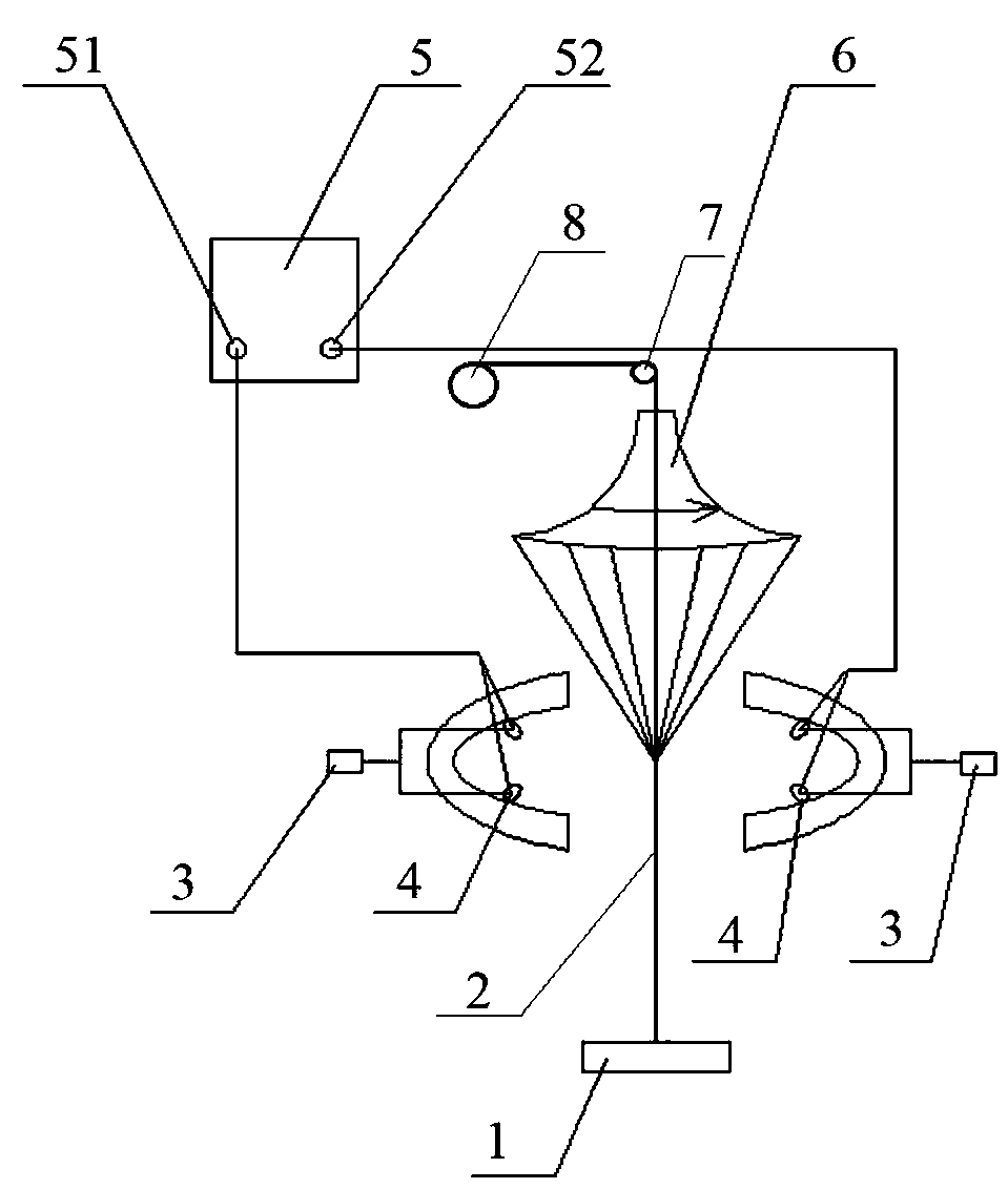
[0026] (2) Build the electrospinning device according to the figure 1 (1 winding ...
Embodiment 2
[0032] A preparation method of multi-layer nanofiber micro-diameter blood vessel tissue engineering scaffold material, the steps are as follows:
[0033] (1) Dissolve the poly(L-lactic acid-polycaprolactone copolymer (PLCL) and silk fibroin (SF) in step (1) in hexafluoroisopropanol (HFIP) at a mass ratio of 10:1, Diol 400 is used as an emulsifier, emulsified to obtain a solution containing heparin and endothelial cell growth factor (VEGF) as the inner layer spinning solution A of the tissue engineering stent material; PLGA and silk fibroin (SF) are placed in a mass ratio of 10:1 In hexafluoroisopropanol (HFIP), after being dissolved evenly, add a certain amount (0.3%) of platelet-derived factor (PDGF) and stir evenly to obtain the middle layer spinning solution B of the stent material; polycaprolactone (PCL) Dissolved in HFIP to obtain a spinning solution C with a mass concentration of 7% of the outer layer of the stent;
[0034] (2) Build an electrospinning device, use nylon f...
Embodiment 3
[0039] A preparation method of multi-layer nanofiber micro-diameter blood vessel tissue engineering scaffold material, the steps are as follows:
[0040] (1) Dissolve the poly(L-lactic acid-polycaprolactone copolymer (PLCL) and silk fibroin (SF) in step (1) in hexafluoroisopropanol (HFIP) at a mass ratio of 1:1, and dissolve them in polyethylene Diol 400 is used as an emulsifier, emulsified to obtain a solution containing heparin and endothelial cell growth factor (VEGF) as the inner layer spinning solution A of the tissue engineering stent material; PLGA and silk fibroin (SF) are placed in a mass ratio of 1:1 In hexafluoroisopropanol (HFIP), after being dissolved evenly, add a certain amount (0.5%) of platelet-derived factor (PDGF) and stir evenly to obtain the middle layer spinning solution B of the stent material; polycaprolactone (PCL) Dissolved in HFIP to obtain a spinning solution C with a mass concentration of 10% of the outer layer of the stent;
[0041] (2) Build an ...
PUM
| Property | Measurement | Unit |
|---|---|---|
| thickness | aaaaa | aaaaa |
| thickness | aaaaa | aaaaa |
| diameter | aaaaa | aaaaa |
Abstract
Description
Claims
Application Information
 - R&D
- Intellectual Property
- Life Sciences
- Materials
- Tech Scout
- Unparalleled Data Quality
- Higher Quality Content
- 60% Fewer Hallucinations
Browse by: Latest US Patents, China's latest patents, Technical Efficacy Thesaurus, Application Domain, Technology Topic, Popular Technical Reports.
© 2025 PatSnap. All rights reserved.Legal|Privacy policy|Modern Slavery Act Transparency Statement|Sitemap|About US| Contact US: help@patsnap.com


