Establishment method of lung cancer diagnosis model

A construction method and a technology for lung cancer diagnosis, which are applied in the preparation, sampling, and measuring devices of test samples, can solve the problems that the diagnostic accuracy and sensitivity need to be improved, the success rate of biopsy is not high, and the prognosis of tumors cannot be judged, etc. Achieve rich biochemical information, wide detection coverage and high sensitivity

Inactive Publication Date: 2017-08-18
中国人民解放军第三0七医院
View PDF0 Cites 5 Cited by
  • Summary
  • Abstract
  • Description
  • Claims
  • Application Information

AI Technical Summary

Problems solved by technology

At present, the examination methods clinically used for the diagnosis of lung cancer mainly include CT, PET-CT, bronchoscopy, exfoliative cytology examination, tissue biopsy, etc., but the cost is high, the success rate of biopsy is not high, and the follow-up period must be followed up. Due to the disadvantage of receiving multiple high doses of ray radiation, the diagnostic accuracy and sensitivity also need to be improved, and the prognosis of the tumor cannot be judged. Therefore, it is particularly necessary to explore markers related to lung cancer diagnosis, treatment effect follow-up, and prognosis

Method used


Image

  • Establishment method of lung cancer diagnosis model
  • Establishment method of lung cancer diagnosis model
  • Establishment method of lung cancer diagnosis model

Examples


Embodiment 1

[0033] The construction of embodiment 1 lung cancer diagnosis model

[0034] 1. Obtain tissue specimen slices

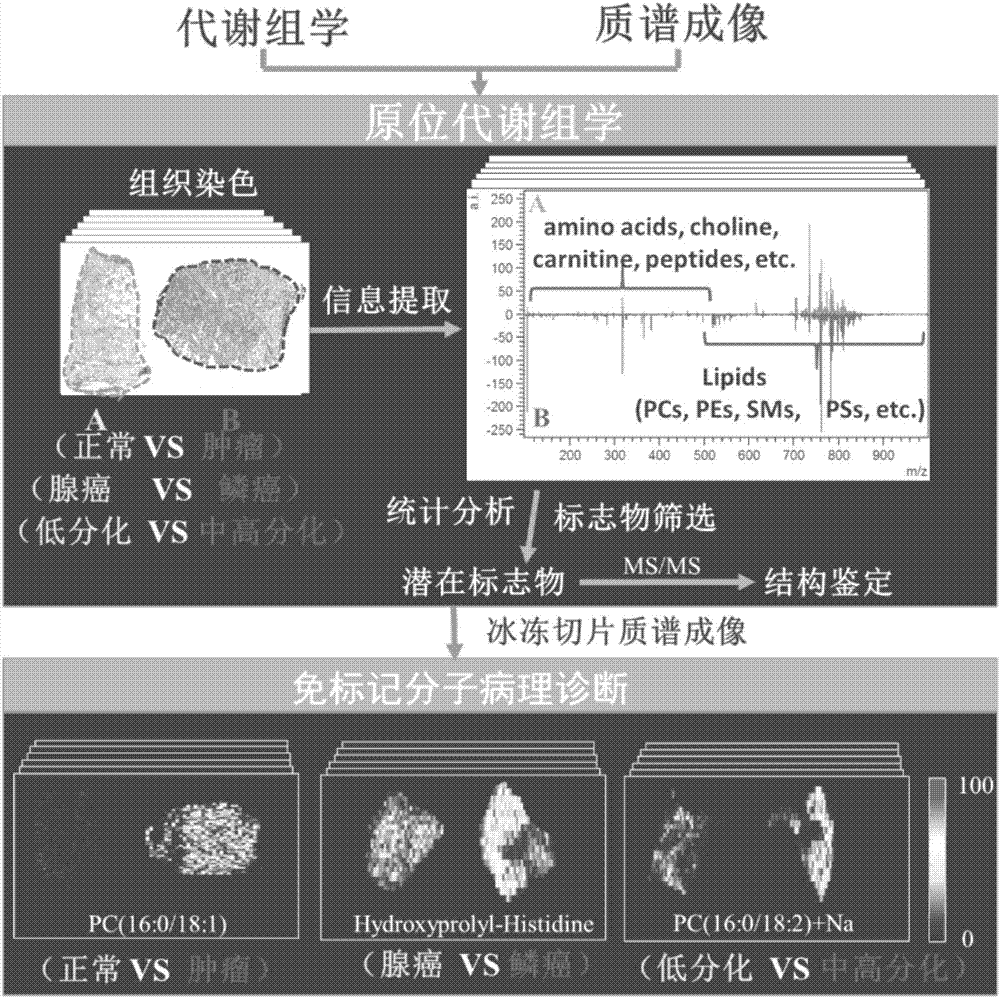
[0035] Three groups of fresh and frozen tissue samples were removed by surgery, including: 1) lung cancer tissue, normal tissue, paracancerous tissue, 2) adenocarcinoma tissue, squamous cell carcinoma tissue, small cell carcinoma tissue, 3) EGFR gene mutation and EGFR wild-type lung cancer organize.

[0036] The slices of the tissue specimens of the 3 groups were prepared to have a thickness of 8um and stained with H&E.

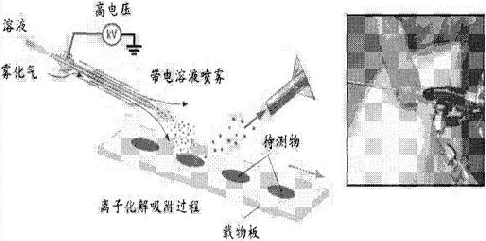
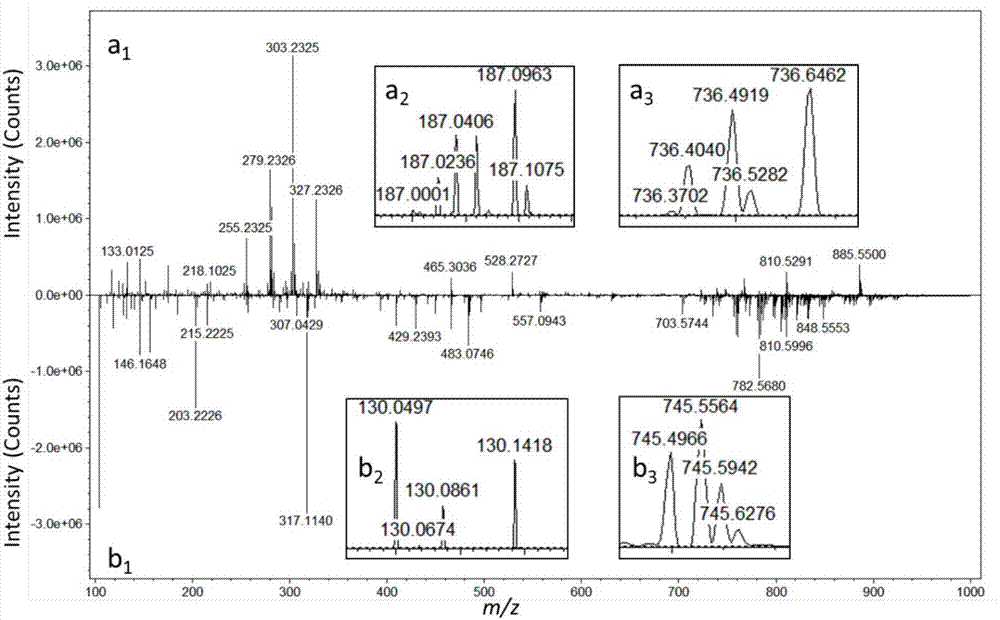
[0037] 2. AFAI-MSI of tissue slices (principle such as figure 1 shown) scanning and data processing

[0038] AFAI-MSI analysis of sections of tissue specimens from 3 groups was performed on a Q Exactive hybrid quadrupole Orbitrap mass spectrometer (ThermoFisher Scientific, USA) equipped with a custom AFAI ion source, acquiring data in positive and negative ion modes. In the positive ion scan mode, methanol and water (8:2, v / v) were mixed with 0....


PUM


Abstract

The invention discloses an establishment method of a lung cancer diagnosis model. The method comprises steps as follows: (1) three groups of tissue sample slices are obtained; (2) a metabolism lipid molecule distribution map of the three groups of tissue sample slices is acquired through AFAI-MS (air flow assisted ionization-mass spectrometry), lipid molecule expression difference of different tissues in the groups is sought, and a rapid lung cancer diagnosis model, a lung cancer molecule pathology diagnosis model and a lung cancer gene diagnosis model for rapidly distinguishing cancer and normal tissue are established; the three groups of tissue sample slices comprise 1) lung cancer tissue, normal tissue and adjacent tissue; 2) adenocarcinoma tissue, squamous carcinoma tissue and small cell carcinoma tissue; 3) EGFR gene mutation and EGFR wild type lung cancer tissue. The lung cancer diagnosis model can more quickly, sensitively and specifically screen lung cancer, and further, lung cancer treatment is effectively guided.

Description

technical field [0001] The invention relates to the field of molecular biology analysis and visualization instrument development. More specifically, it relates to a method for constructing a lung cancer diagnosis model. Background technique [0002] Mass spectrometry imaging is a new molecular imaging technology. It can obtain the spatial distribution of molecules in tissue slices. Compared with the existing technology, it does not require specific labels. Through one imaging scan analysis, hundreds of different molecules can be obtained. distribution map. Mass spectrometry imaging technology is expected to become an ideal histopathological examination method through the correlation of multi-analyte distribution maps with histopathological or clinical information. For example, MALDI-MSI imaging technology can detect compounds such as peptides, proteins, carbohydrates, and lipids, and its detection limit is within the range of atto molar levels. The spatial resolution of m...

Claims


Application Information

Patent Timeline
Patent Type & Authority Applications(China)
IPC IPC(8): G01N27/62G01N1/28G01N1/30
CPCG01N27/62G01N1/286G01N1/30G01N2001/2873
Inventor 章敏
Owner 中国人民解放军第三0七医院
Why Eureka
Try Eureka