Serum or plasma microRNA as biomarkers for non-small cell lung cancer

a lung cancer and serum microrna technology, applied in the field of serum or plasma microrna as biomarkers for non-small cell lung cancer, can solve the problems of insufficient early stage diagnosis of cancer, inability to use detection results as accurate indicators of disease diagnosis, and inability to accurately diagnose diseas

Active Publication Date: 2016-07-12
MICROMEDMARK BIOTECH CO LTD
View PDF13 Cites 0 Cited by
  • Summary
  • Abstract
  • Description
  • Claims
  • Application Information

AI Technical Summary

Benefits of technology

[0062](1) The methods using screened microRNAs in serum or plasma as the markers of non-small cell lung cancer possess advantages including wide detection coverage, high sensitivity, low cost, convenient sample collection and preservation (serum or plasma can be preserved at −20° C.), among others. The method can be widely applied in general disease investigation and become an effective means for early diagnosis.
[0063](2) Having serum or plasma microRNAs as novel disease markers can solve the problems of low specificity and sensitivity in signal marker detection method caused by unavoidable individual differences, will markedly improve the clinical detectability of diseases and achieve early diagnosis and treatment.
[0064](3) The novel serum or plasma microRNA detection technique enable detection of a series of disease related markers, so that the influence from the individual differences of the patients (including age, gender, race, diet and environment conditions, and etc.) can be avoided, which is one of the major problems that single marker detection techniques cannot solve.
[0065]In summary, the present invention can be further applied in the early stage diagnosis of non-small cell lung cancer, and the novel markers of non-small cell lung cancer in serum or plasma do not only provide the foundation for understanding the mechanism of non-small cell lung cancer more fully at the molecular level, but also accelerate the advances in clinical diagnosis and treatment. It is quite apparent that, with the advantages of the serum or plasma microRNAs, the serum or plasma microRNAs diagnosis technique for severe diseases including cancer shall become part of general physical examination; as the genetic treatment related to microRNAs is being widely applied, these diseases shall be conquered.

Problems solved by technology

Although many disease markers have been discovered and applied in the general survey and diagnosis of clinical diseases and in the monitoring of therapeutic efficacy, obvious deficiencies exist in their clinical application.
For instance, the tumor markers that have been widely used in clinical diagnosis, including alpha-fetoprotein (AFP), lactic dehydrogenase (LHD) and carcinoembryonic antigen (CEA), are inadequate for the early stage diagnosis of cancers for various reasons.
For example, the sensitivity and specificity of the disease markers mentioned above are relatively low, so that the detection results cannot be used as accurate indicators for disease diagnosis.
Furthermore, the disease markers mentioned above cannot satisfy the requirement for early stage diagnosis, which often bears a positive correlation with the therapeutic efficacy.
Due to drawbacks including the extremely high specificity of cancer differentiation types, the relatively low sensitivity of tumor bodies, the difficulty of taking repetitive samples, the high requirements for preserving samples, and the expensive costs, the application of the currently known tumor markers cannot be widely promoted under present conditions.
Meanwhile, some conventional medical methods such as biopsy for tissue and cell detection have inherent deficiencies, such as taking sample from an incorrect location, insufficiency of tissue and cell sample materials, or the technician's lack of experience, which may all contribute to misdiagnosis.
Other techniques like medical imaging that have been widely used in disease inspection and diagnosis are rather limited in the qualitative description of disease process.
However, the wide clinical application is restrained by difficulties in obtaining tissue samples.

Method used


Image

  • Serum or plasma microRNA as biomarkers for non-small cell lung cancer
  • Serum or plasma microRNA as biomarkers for non-small cell lung cancer
  • Serum or plasma microRNA as biomarkers for non-small cell lung cancer

Examples


example 1

RT-PCR Experiments of microRNAs in Serum or Plasma

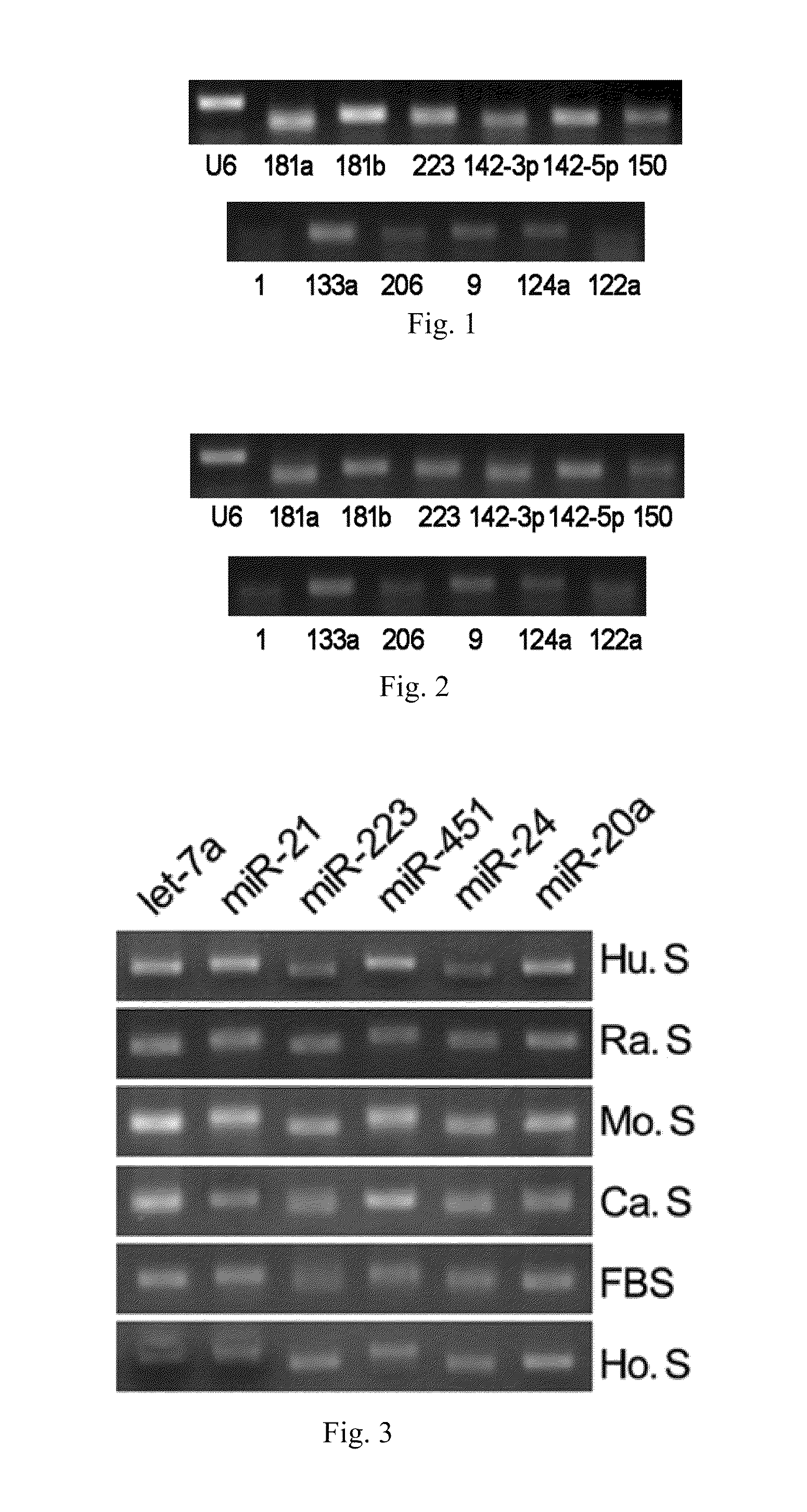
[0067]Using the RT-PCR assay, it has been shown that various microRNAs exist stably in serum or plasma of both human beings and other animals, and that their expression levels are considerably high. The specific steps are as follows:[0068](1) Collecting serum or plasma of mice, rats, normal subjects and patients with certain diseases.[0069](2) Preparing cDNA samples. This operation has two options: one is to directly conduct the reverse transcription reaction with 10 μl serum or plasma; the other is to firstly extract the total RNA from serum or plasma (usually, about 10 μg of RNA can be enriched by 10 μl of serum or plasma) with Trizol reagent (Invitrogen Co.), and then to obtain cDNA by the RNA reverse transcription reaction. The reaction system of reverse transcription comprises 4 μl of 5×AMV buffer, 2 μl, 10 mM of each dNTP mixture (Takara Co.), 0.5 μl of RNase Inhibitor (Takara Co.), 2 μl of AMV (Takara Co.) and 1.5 μl of gene s...

example 2

Real-Time PCR Experiments of microRNAs in Serum or Plasma

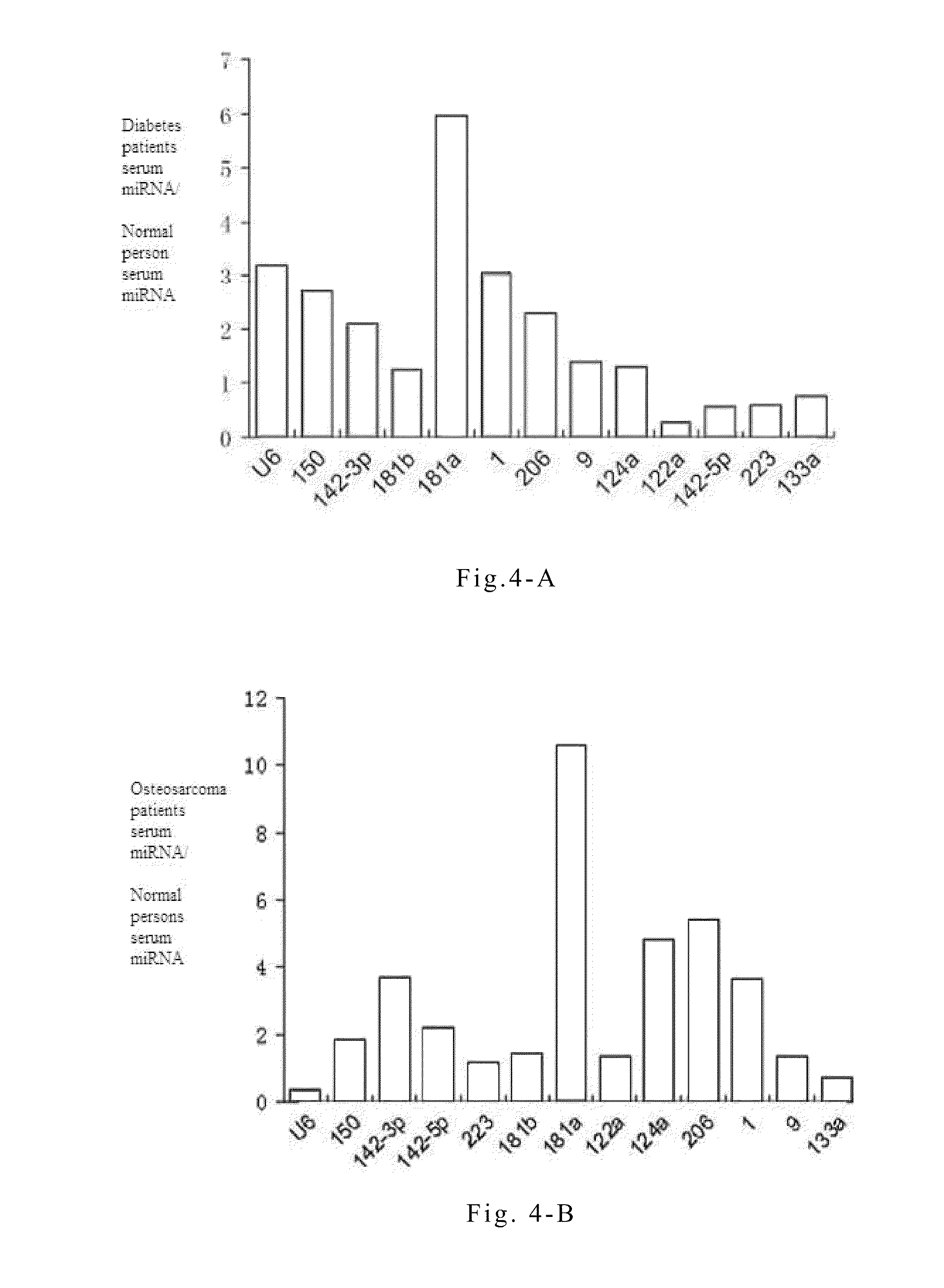
[0073]Quantitative PCR experiments on serum or plasma microRNAs are conducted in order to study the specific variations of these microRNAs during the progression of non-small cell lung cancer. The principles and steps of the quantitative experiment are the same as that of RT-PCR, except that the fluorescent dye EVA GREEN is added during PCR. An ABI Prism 7300 fluorescent quantitative PCR system is used to conduct PCR reaction under the following conditions: one cycle at 95° C. for 5 mins followed by 40 cycles at 95° C. for 15 seconds and 60° C. for 1 minute. The data processing method is the ΔΔCT method, wherein CT is the number of cycles when the reaction reaches the threshold. The expression level of each microRNA relative to an internal standard reference can be expressed by the equation 2−ΔCT, wherein ΔCT=CTsample−CTinternal reference. The reverse transcription reactions are directly conducted on serum or plasma samples of...

example 3

Biochip Utilizing Serum or Plasma MicroRNAs for the Diagnosis of Non-Small Cell Lung Cancer

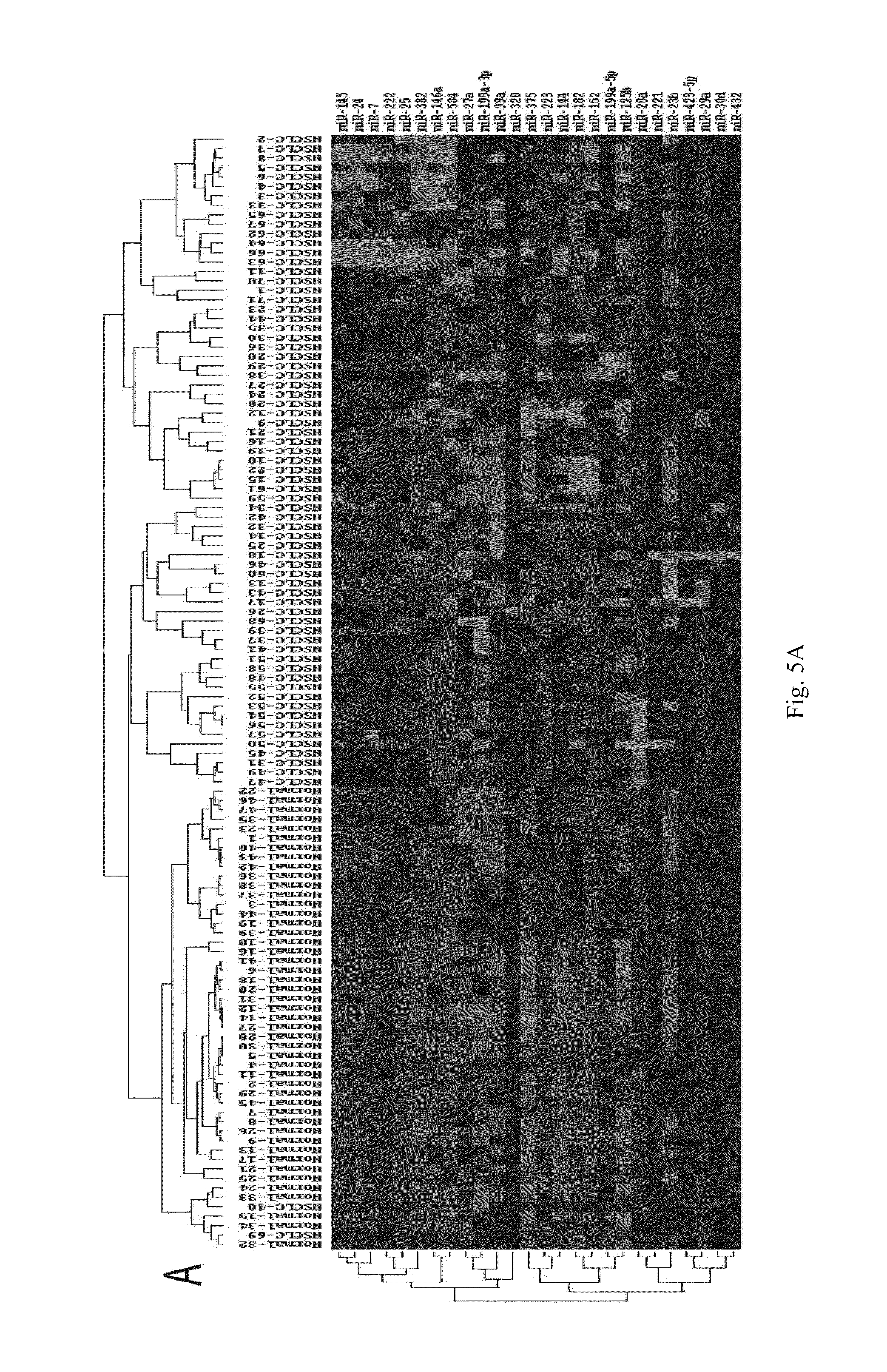
[0075]The operation steps of the biochip are as follows:[0076](1) Extracting the total RNA from serum or plasma, and measuring the mass of total RNA by formaldehyde denaturing gel electrophoresis.[0077](2) Separating microRNAs: microRNAs are separated with Ambion's microRNA Isolation Kit (Cat #.1560) from 50-100 m of total RNA.[0078](3) Conducting the fluorescent labeling on microRNA samples: the microRNA samples are labeled with fluorescent labeling using the T4RNA ligase labeling method, then precipitated using absolute ethanol, and then dried before hybridization to the biochip.[0079](4) Hybridizing and rinsing: RNAs are dissolved in 16 μl of hybridizing solution (15% formamide; 0.2% SDA; 3×SSC; 50×Denhardt's solution), hybridized overnight at 42° C. After hybridization, the RNAs are rinsed for 4 mins in a solution of 0.2% SDS, 2×SSC at about 42° C., and for 4 mins in the solution of 0.2% S...


PUM

PropertyMeasurementUnit
medical imagingaaaaaaaaaa
lengthaaaaaaaaaa
resistanceaaaaaaaaaa

Abstract

The present invention provides non-small cell lung cancer markers and the use thereof in diagnosing and monitoring diseases in vitro. The non-small cell lung cancer markers include at least one of the 26 selected detectable mature microRNAs existing stably in human serum or plasma. The invention also provides probe combinations, a kit and biochip for detecting the non-small cell lung cancer markers. The invention further provides a method for detecting the said lung cancer markers. The method in the present invention enables extensive detection spectrum, high sensitivity, low cost, convenient sample taking and preservation. The method can be applied in the general survey of disease, solves problems of the low specificity and sensitivity encountered with previous single markers, and increases significantly the clinical detection rate of diseases; it forms an effective means for diagnosing diseases at an early stage.

Description

RELATED APPLICATIONS[0001]This application is a U.S. National Phase of International Application No.: PCT / CN2009 / 001441, filed Dec. 14, 2009, designating the U.S. and published on June 24, 2010 as WO2010 / 069129A1, which claims the benefit of Chinese Application No. 200810243501.7, filed Dec. 15, 2008.BACKGROUND OF THE INVENTION[0002]The present invention relates to biological diagnostic techniques, and more specifically to the separation and the qualitative and quantitative analysis of microRNAs in human serum or plasma, and to clinical indicators of non-small cell lung cancer. More particularly, the invention relates to a method for detecting and analyzing variations in microRNAs in human serum or plasma. This enables in vitro diagnosis of non-small cell lung cancer; evaluation of disease stage; monitoring of the progression of non-small cell lung cancer; evaluation of disease complications and relapse; prognosis of non-small cell lung cancer; and evaluation of drug efficacy and th...

Claims


Application Information

Patent Timeline
Patent Type & Authority Patents(United States)
IPC IPC(8): C12Q1/68
CPCC12Q1/6886C12Q2600/112C12Q2600/118C12Q2600/158C12Q2600/178C12Q2600/16
Inventor ZHANG, CHENYUZENG, KEZHANG, JUNFENGBA, YICHEN, XILI, HAIJIN
Owner MICROMEDMARK BIOTECH CO LTD
Why Eureka
Try Eureka