Nucleic acid aptamer-polypeptide complex probe and its preparation method and application

A nucleic acid aptamer and polypeptide complex technology, which is applied in the fields of biology and medicine, can solve problems such as low solubility and low enzymatic hydrolysis efficiency, and achieve high affinity, high purity, and simple and easy preparation methods

Active Publication Date: 2020-12-01
NANJING MEDICAL UNIV
View PDF2 Cites 0 Cited by
  • Summary
  • Abstract
  • Description
  • Claims
  • Application Information

AI Technical Summary

Problems solved by technology

However, the direct application of conventional directed proteomics methods to quantify transmembrane proteins such as HER2 protein will encounter some special difficulties, mainly due to the hydrophobicity of membrane proteins and the natural resistance to proteolysis, in addition to low Solubility and low enzymatic efficiency may be another confounding factor

Method used


Image

  • Nucleic acid aptamer-polypeptide complex probe and its preparation method and application
  • Nucleic acid aptamer-polypeptide complex probe and its preparation method and application
  • Nucleic acid aptamer-polypeptide complex probe and its preparation method and application

Examples


Embodiment 1

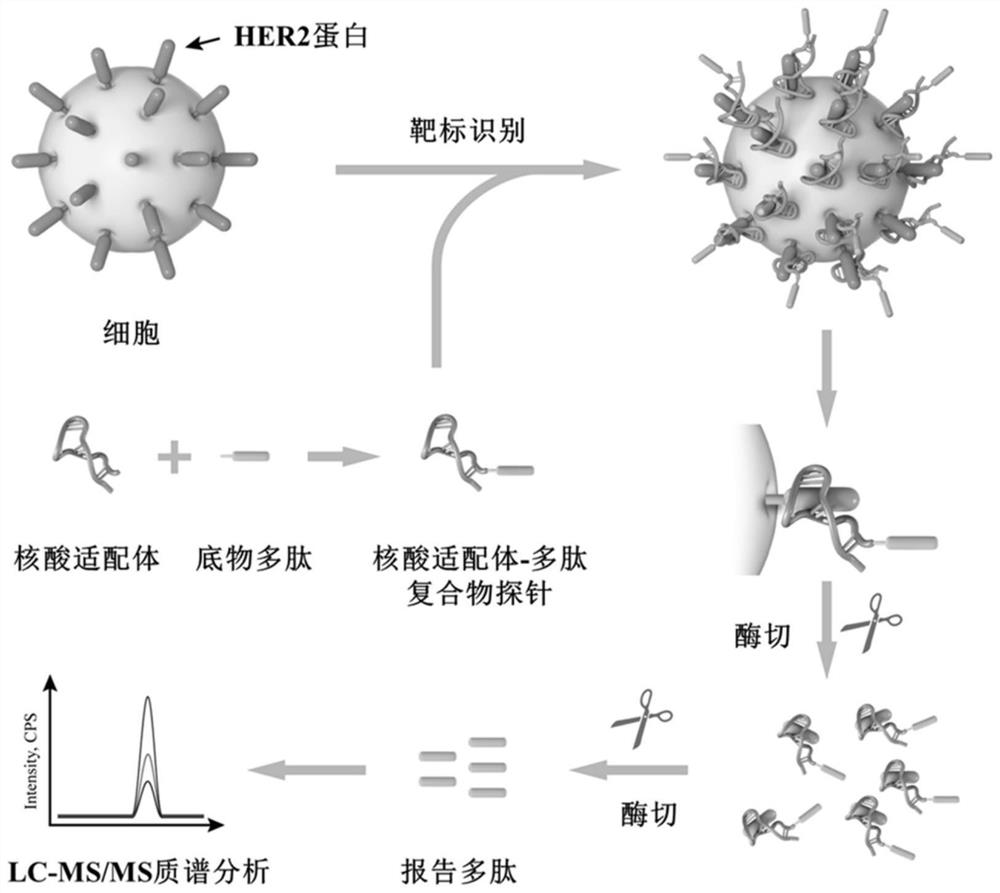
[0035] Synthesis and identification of embodiment 1 nucleic acid aptamer-polypeptide complex probe

[0036] (1) Preparation and purification of nucleic acid aptamer-polypeptide complex probes

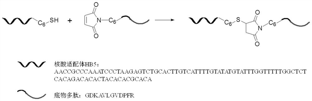
[0037] With trichloroethyl phosphate (TCEP) as reducing agent, 100 μL of disulfide bond-modified nucleic acid aptamer HB5 at a concentration of 20 μM

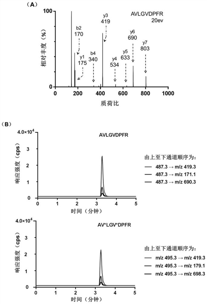
[0038] (AACCGCCCAAATCCCTAAGAGTCTGCACTTGTCATTTTGTATATGTATTTGGTTTTTGGCTCTCACAGACACACTACACACGCACA) was mixed with 50 μL of TCEP-reduced beads, and reacted and shaken at 37°C for 2h. The samples were then centrifuged at 1000 x g for 5 min. Take the supernatant containing reduced HB5, add the same volume of 200 μM maleimide-modified substrate polypeptide (GDKAVLGVDPFR) to the supernatant, shake vigorously at 37°C for 4 hours to carry out the conjugation reaction, and then use conjugation The difference in retention time before and after the yoke (the retention times of the nucleic acid aptamer and the nucleic acid aptamer-polypeptide comple...

Embodiment 2

[0046] Example 2 Detection of HER2 protein content on the surface of breast cancer cells with nucleic acid aptamer-polypeptide complex probe

[0047] In order to prevent damage to the proteins on the cell surface, we collected HER2-positive breast cancer cells (BT474 and SK-BR-3) and HER2-negative breast cancer cells (MDA-MB-231 and MCF -7). After the cells were washed three times with PBS, they were blocked with 200 μL of binding buffer at a blocking temperature of 37° C. for 30 min. Then at a cell number of about 1 x 10 6 200 μL of the probe with a concentration of 1 μM was added to the cells, and the mixture was shaken and reacted at 37° C. for 2.5 hours. After the reaction, the cells were washed three times with 500 μL of cold PBS to remove unbound probes, and then washed twice with 100 μL of 50 mM ammonium bicarbonate solution. Then surface enzyme digestion (or restriction enzyme digestion) technology is mainly used to release the reporter polypeptide from the biologic...

Embodiment 3

[0050] Example 3 Detection of HER2 protein content on breast cancer tissue sections with nucleic acid aptamer-polypeptide complex probe

[0051]36 pairs of human breast primary tumor tissues and their corresponding paracancerous tissues were cut into 6 μm tissue sections on a frozen microtome treated with 100% alcohol and 100% methanol, and 200 μL of binding buffer was dropped on the tissues On the slice, shake gently at 37°C for 1 h, and then wash the tissue slice three times with binding buffer. Then, 200 μL of binding buffer containing 200 pmol probe was added and incubated at 37° C. for 2.5 h. Finally, the tissue sections were washed three times with PBS and twice with 50 mM ammonium bicarbonate solution. Incubate the probe-loaded sections with trypsin, and collect the post-reaction solution in a centrifuge tube. After centrifugation at 20,000×g at 4°C for 10 min, 2 μg of trypsin was added to the centrifuge tube, and then reacted at 37°C for 24 hours, and 10 μL of 0.1% t...


PUM


Abstract

The invention discloses an aptamer-polypeptide compound probe as well as a preparation method and application thereof. The aptamer-polypeptide compound probe has a following structure, and the structural formula is as shown in the specification, wherein R1 is an aptamer HB5, and the R2 is a substrate polypeptide. According to the aptamer-polypeptide compound probe disclosed by the invention, the aptamer technology is combined with a targeted proteomics technology, high specificity and high affinity of an aptamer recognized target are combined with high sensitivity, high selectivity and wide dynamic range detected by the targeted proteomics technology, and a brand new quasi-targeted proteomics method for quantitative HER2 proteins is developed.

Description

technical field [0001] The invention belongs to the technical field of biology and medicine, and specifically relates to a nucleic acid aptamer-polypeptide complex probe and its preparation method and application. Background technique [0002] HER2 (also known as Neu, ErbB-2, CD340, and p185) is a transmembrane receptor protein that is overexpressed in HER2-positive breast cancer cells. Overexpression of HER2 can enhance the invasiveness, viability and regeneration ability of cancer cells. So far, many evidences have shown that early diagnosis and timely supplementary treatment (such as Herceptin (trastuzumab)) can well control the development of HER2-positive breast cancer. [0003] Some existing techniques such as Western Blotting, enzyme-linked immunosorbent assay (ELISA), immunohistochemistry (IHC), fluorescence in situ hybridization (FISH), etc. can detect the expression of HER2 in breast cancer, among which The detection of HER2 protein overexpression by immunohistoc...

Claims


Application Information

Patent Timeline
Patent Type & Authority Patents(China)
IPC IPC(8): C07K7/08G01N33/68G01N33/574
CPCC07K7/08C07K19/00G01N33/57415G01N33/68G01N2333/71
Inventor 陈芸许飞飞周伟贤
Owner NANJING MEDICAL UNIV
Why Eureka
Try Eureka