Two-photon fluorescence microscopy method and device based on photon recombination

A two-photon fluorescence and photon recombination technology, applied in the field of confocal microscopy, can solve problems such as high alignment accuracy, and achieve the effects of high signal-to-noise ratio, large imaging depth and simple structure

Inactive Publication Date: 2017-12-15
ZHEJIANG UNIV
View PDF4 Cites 6 Cited by
  • Summary
  • Abstract
  • Description
  • Claims
  • Application Information

AI Technical Summary

Problems solved by technology

However, structured light microscopy is generally used in wide-field microscopy. Although stimulated emission depletion microscopy can be applied to two-photon microscopy, it requires high alignment accuracy.

Method used


Image

  • Two-photon fluorescence microscopy method and device based on photon recombination
  • Two-photon fluorescence microscopy method and device based on photon recombination
  • Two-photon fluorescence microscopy method and device based on photon recombination

Examples


Embodiment 1

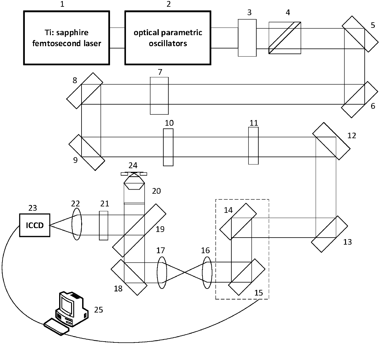
[0054] Such as figure 2 As shown, a virtual fluorescence differential microscopy device based on photon recombination to realize sample scanning by galvanometer scanning, including femtosecond laser 1, optical parametric oscillator 2, half-wave plate 3, polarization beam splitter 4, mirror 5, Reflector 6, half-wave plate 7, reflector 8, reflector 9, quarter-wave plate 10, quarter-wave plate 11, reflector 12, reflector 13, scanning galvanometer 14, scanning galvanometer 15 , scanning mirror 16, field lens 17, mirror 18, dichroic mirror 19, microscope objective lens 20, filter 21 and field lens 22.

[0055] use figure 2 The virtual fluorescence differential microscopy method implemented by the device shown is as follows:

[0056] (1) Femtosecond laser 1 emits illuminating light, exits through optical parametric oscillator 2, becomes s-light, p-light ratio modulated polarized light through half-wave plate 3, realizes for Adjustment of light intensity in the light path;

[0...

Embodiment 2

[0062] Such as Figure 4 As shown, a two-photon microscopy device based on photon recombination that realizes sample scanning by a nano-translation stage, including a femtosecond laser 1, an optical parametric oscillator 2, a half-wave plate 3, a polarization beam splitter 4, and a mirror 5, mirror 6, mirror 8, mirror 9, mirror 12, mirror 13, mirror 14, half-wave plate 7, quarter-wave plate 10, quarter-wave plate 11, dichroic mirror 15, Optical filter 17, field lens 18, array detector 19, nano translation stage 21, computer 22.

[0063] use Figure 4 The photon-sufficient two-photon microscopy method realized by the setup shown is as follows:

[0064] (1) The femtosecond laser 1 emits ultrashort pulse light, passes through the optical parametric oscillator 2, passes through the half-wave plate 3 and the polarizing beam splitter 4, and obtains a linearly polarized light beam whose intensity can be modulated;

[0065] (2) the linearly polarized light is incident on the half-w...


PUM


Abstract

The invention discloses a two-photon fluorescence microscopy device based on photon recombination. The device comprises a femtosecond laser device, an optical parameter oscillator, a first half wave plate, a polarization beam splitter, a second half wave plate, a first quarter wave plate, an objective lens, an array detector and a computer, wherein the femtosecond laser device is used for sending ultra-short pulse laser to perform two-photon excitation on samples; the first half wave plate is used for regulating the proportion of light s and light p of emergent light of the optical parameter oscillator; the polarization beam splitter is used for regulating the light intensity; the second half wave plate is used for regulating the polarization direction of the line polarization; the first quarter wave plate is used for converting the line polarization light into round polarization light; the objective lens is used for focusing the round polarization light into solid round spots for illuminating a sample and exciting the fluorescence; the array detector consists of a plurality of detector units, and is used for collecting fluorescence signals; the computer processes the fluorescence signals obtained by each detector unit to obtain an image corresponding to one object point. The invention also discloses a two-photon fluorescence microscopy method based on photon recombination. The detector array is used for replacing the original single needle hole detector, so that the obtained image has a higher signal-to-noise ratio.

Description

technical field [0001] The invention belongs to the field of confocal microscopy, in particular to a two-photon fluorescence microscopy method and device based on photon reconstruction. Background technique [0002] Two-photon fluorescence microscopy enables three-dimensional imaging in living biological samples. Compared with traditional confocal microscopy, two-photon can achieve better optical sectioning function, deeper penetration depth and less phototoxicity. The single-photon excitation process is that the ground state electron absorbs a short-wavelength photon to reach the excited state. This process generally requires ultraviolet or blue / green light, and these short-wavelength light may cause severe phototoxicity to biological samples. For two-photon excitation, under the condition of sufficient light intensity, the excitation of ground state electrons can be realized by simultaneously absorbing two photons with lower energy (the wavelength is usually in the infra...

Claims


Application Information

Patent Timeline
Patent Type & Authority Applications(China)
IPC IPC(8): G01N21/64G01N21/01
CPCG01N21/6402G01N21/01
Inventor 匡翠方孙试翼刘旭李海峰徐良毛磊张克奇
Owner ZHEJIANG UNIV
Why Eureka
Try Eureka