Cardiac muscle cell detection method based on inverse opal structure hydrogel and application thereof

A technology of anti-opal structure and cardiomyocytes, applied in the field of biomedical material research, to achieve the effect of easy-to-obtain materials, easy large-scale production, and intuitive effects

Active Publication Date: 2018-02-02
SOUTHEAST UNIV
View PDF14 Cites 13 Cited by
  • Summary
  • Abstract
  • Description
  • Claims
  • Application Information

AI Technical Summary

Problems solved by technology

Therefore, the research and development of sensing materials with high sensitivity to detect the contractility and beating frequency of cardiomyocyt

Method used


Image

  • Cardiac muscle cell detection method based on inverse opal structure hydrogel and application thereof
  • Cardiac muscle cell detection method based on inverse opal structure hydrogel and application thereof

Examples


Embodiment 1

[0038] Embodiment 1 A kind of cardiomyocyte detection and application based on gelatin methacrylate (GelMA) inverse opal structure hydrogel

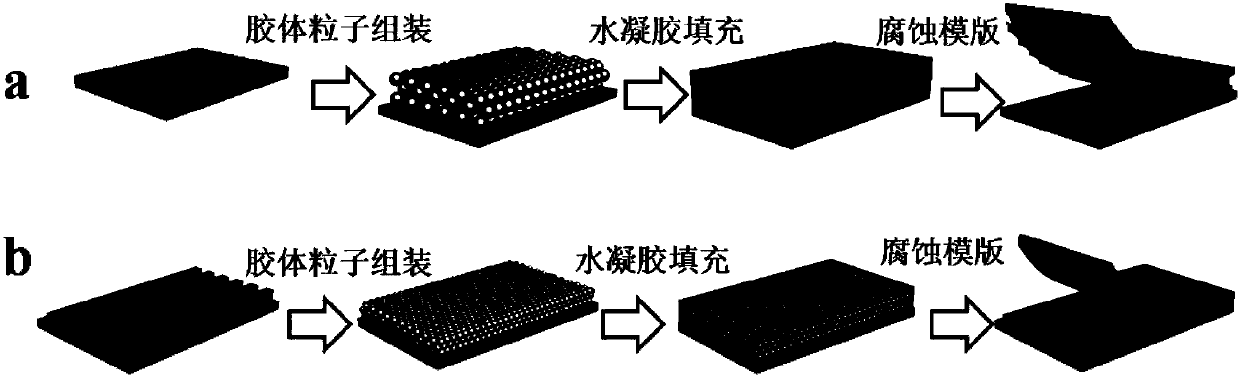
[0039] 1. Preparation of GelMA inverse opal hydrogel:

[0040] 1) Disperse the purified monodisperse silicon dioxide particles with a particle size of 300nm in an ethanol solution to obtain a silicon dioxide ethanol dispersion with a concentration of silicon dioxide particles of 20wt%;

[0041] 2) Deposit the silicon dioxide ethanol dispersion on the glass sheet to form a silicon dioxide photonic crystal template, and finally perform high-temperature calcination (500° C.) on the obtained photonic crystal template to obtain a photonic crystal template with better mechanical strength;

[0042] 3) Soak the photonic crystal template with better mechanical strength in the GelMA hydrogel pre-poly solution (0.3g / ml) for 2 hours, and obtain the photonic crystal-hydrogel hybrid system through UV curing;

[0043] 4) Finally, HF (2wt%) was used to...

example 2

[0052] Example 2 A kind of cardiomyocyte detection based on bovine serum albumin (BSA) inverse opal hydrogel membrane and its application

[0053] 1. Preparation of BSA inverse opal structure hydrogel film:

[0054] 1) Dispersing the purified polystyrene particles monodispersed at 100 nm in an aqueous solution to obtain a polystyrene aqueous solution with a concentration of 1 wt %;

[0055] 2) Deposit polystyrene aqueous solution on the silicon wafer, self-assemble to form polystyrene photonic crystal template;

[0056] 3) Soak the polystyrene photonic crystal template in BSA hydrogel prepolymer solution (0.2g / ml) for 3 hours, and cross-link with glutaraldehyde to obtain a photonic crystal-hydrogel hybrid system;

[0057] 4) Finally, absolute ethanol was used to dissolve the polystyrene particles in the photonic crystal hydrogel hybrid system to obtain the BSA inverse opal hydrogel.

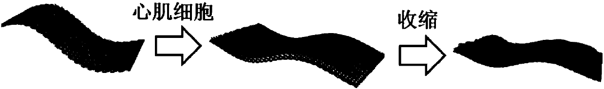
[0058] 2. BSA inverse opal hydrogel for the culture of cardiomyocytes

[0059] The cardiom...

example 3

[0066] Example 3 A kind of cardiomyocyte detection based on gelatin inverse opal structure hydrogel membrane and its application

[0067] 1. Preparation of gelatin stripe inverse opal structure hydrogel:

[0068] 1) Disperse the purified silicon dioxide particles monodispersed at 200nm in an ethanol solution to obtain a silicon dioxide ethanol dispersion with a concentration of silicon dioxide particles of 15% by weight;

[0069] 2) Deposit the silica ethanol dispersion on the striped silicon wafer (concave 40 μm, convex 30 μm) to form a striped silica photonic crystal template, and finally perform high-temperature calcination (600 ° C) on the obtained striped photonic crystal template to obtain mechanical strength Better stripe photonic crystal template;

[0070] 3) Soak the striped photonic crystal template with better mechanical strength in gelatin hydrogel prepolymer solution (0.25g / ml) for 5h, and cross-link with glutaraldehyde (3wt%) to obtain a photonic crystal hydroge...


PUM

PropertyMeasurementUnit
Particle sizeaaaaaaaaaa

Abstract

The invention discloses a cardiac muscle cell detection method based on inverse opal structure hydrogel and application thereof. The detection method comprises the following steps: (1) preparing inverse opal structure hydrogel of biocompatibility; (2) culturing cardiac muscle cells based on inverse opal structure hydrogel; (3) detecting the cardiac muscle cells; (4) analyzing data. The inverse opal structure hydrogel in the invention has good biocompatibility, cells have high activity and phenotype when growing on the surface of the hydrogel, the inverse opal structure hydrogel not only provides a carrier for the growth of the cardiac muscle cells, but also provides a stable optical sensing signal for detecting the cardiac muscle cell shrinking force and hopping frequency. The detection method does not need a complicated detection system, and has the advantages of intuitiveness, high sensitivity, high efficiency and capability of preventing the influence of external conditions; the method can be used for screening and evaluating heart drugs and can screen the drugs by virtue of the variation of the cardiac muscle cell shrinking force and hopping frequency after the drug is added.

Description

technical field [0001] The invention relates to a cardiomyocyte detection method based on an inverse opal structure hydrogel and an application thereof, belonging to the technical field of biomedical material research. Background technique [0002] At present, in the detection process of cardiomyocytes, we mainly rely on in vitro cell culture technology and the construction of in vivo animal models. In the process of new drug research and development, due to the lack of complexity of the body, simple two-dimensional cell culture has problems such as inability to truly simulate the microenvironment in vivo and the complex physiological processes between tissues and organs; in addition, due to the uncertainty of building animal models With its own complexity and some humanitarian factors, the construction of animal models in drug development also faces certain challenges. In order to truly simulate the microenvironment in the body and realize the real simulation of organs in ...

Claims


Application Information

Patent Timeline
IPC IPC(8): G01N15/10
CPCG01N15/10G01N2015/1006
Inventor 赵远锦付繁繁高崴商逸璇王月桐
Owner SOUTHEAST UNIV
Why Eureka
Try Eureka