
Method for inducing mouse fibroblasts into cartilage by adopting small-molecule composition

A technology of fibroblasts, composition, applied in the field of biotechnology and cartilage repair

Active Publication Date: 2018-02-09
ZHEJIANG UNIV
View PDF6 Cites 9 Cited by
  • Summary
  • Abstract
  • Description
  • Claims
  • Application Information

AI Technical Summary

Problems solved by technology

However, for chondrocytes that also do not have the ability to self-repair, there is no research on inducing the transdifferentiation of fibroblasts to chondrocytes without introducing exogenous genes.

Method used


Image

  • Method for inducing mouse fibroblasts into cartilage by adopting small-molecule composition
  • Method for inducing mouse fibroblasts into cartilage by adopting small-molecule composition
  • Method for inducing mouse fibroblasts into cartilage by adopting small-molecule composition

Examples


Embodiment 1

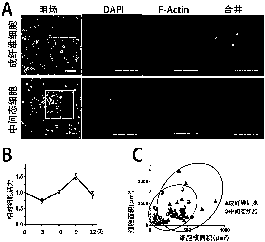
[0053] Example 1: Induction of mouse embryonic fibroblasts into intermediate cells and characterization

[0054] 1. Experiment

[0055] 1.1 Primary extraction and culture of mouse embryonic fibroblasts

[0056] Get 13.5-day-old C57BL / 6J mouse embryos, cut off the head, limbs, internal organs, gonads and spine, mince the remaining tissues, and digest with 0.05% trypsin (Gibico) for 5 minutes. Add high glucose DMEM medium (i.e. H-DMEM medium containing 10% FBS) containing volume concentration 10% fetal bovine serum (FBS, Gibico) to stop digestion, centrifuge at 1200-1500rpm for 3-5 minutes, and precipitate with 10% Resuspend in H-DMEM medium with FBS, put into culture dish, culture at 37°C, replace with fresh H-DMEM medium containing 10% FBS after adherence. Cultivate at 37°C until the cells cover 80-90% of the culture dish, then cryopreserve or subculture, and obtain 1-3 generations of wild-type mouse embryonic fibroblasts.

[0057] 1.2 Chemical induction of intermediate cel...

Embodiment 2

[0083] Example 2: Chondrogenic induction of intermediate cells

[0084] 1. Experiment

[0085] 1.1 Chondrogenic induction of intermediate cells

[0086] The 2nd generation wild-type mouse embryonic fibroblasts prepared in Example 1 were mixed with 1×10 5 The density of cells / well was planted in a 12-well plate supplemented with H-DMEM medium containing 10% FBS, cultivated at 37°C until the cells grew to 90% density, the medium was removed, and the small molecule composition (V0. 5mM+C3μM+R1μM) chemical induction medium, cultivated in 37°C, 5% oxygen, 5% carbon dioxide (the rest is nitrogen) environment for 6-9 days, and replace fresh chemical containing small molecule composition every 2-3 days induction medium. Discard supernatant culture medium, add chondrogenic induction medium, at 37 ℃, 21% oxygen, 5% carbon dioxide (the rest is nitrogen) environment and cultivate 14 days, every 3-4 days change fresh chondrogenic induction medium.

[0087] The final concentration of th...

Embodiment 3

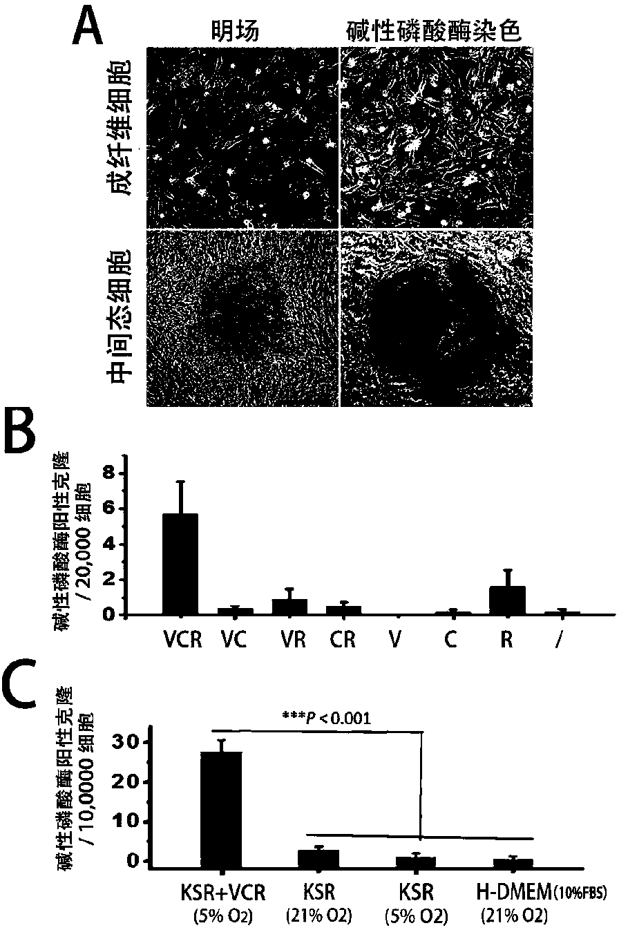
[0108] Example 3: Combined screening to obtain an optimized composition of small molecule compositions

[0109] 1.1 Safranin O staining confirmed the basic induction model.

[0110] The second-generation wild-type mouse embryonic fibroblasts prepared in step 1.1 of Example 1 were planted at a density of 3000 / well in a 96-well plate supplemented with H-DMEM medium containing 10% FBS, and cultured at 37°C until Cells grew to 90% density, the culture medium was removed, slowly added chemical induction medium containing small molecule composition (VPA 0.5mM, CHIR-98014 3μM and Repsox 1μM), 37°C, 5% oxygen, 5% carbon dioxide (other Cultivate in a nitrogen) environment, replace the fresh chemical induction medium containing the small molecule composition VCR every 2-3 days, and after cultivating for 6 days, replace the chemical induction medium as the cartilage induction medium (the composition is the same as that of Example 2 step 1.1) , at 37° C., in an environment of 21% oxygen ...


PUM


Abstract

The invention discloses a method for inducing mouse fibroblasts into cartilage by adopting a small-molecule composition. The method comprises the following steps: carrying out adherent culture on mouse embryo fibroblasts, removing a culture medium, slowly adding a chemical induction culture medium containing the small-molecule composition, carrying out culturing in the environment having the temperature of 37 DEG C and containing 3-8% of oxygen, 3-8 % of carbon dioxide and the balance of nitrogen, wherein the chemical induction culture medium containing the small-molecule composition is replaced every 2-3 days; carrying out continuous culturing for 4-12 days, thus obtaining intermediate state cells, wherein the small-molecule composition comprises an HDAC inhibitor, a GSK-3 inhibitor and aTGF-beta signal channel inhibitor; and transferring the intermediate state cells to a cartilage inducing medium, carrying out culturing in the environment having the temperature of 37 DEG C and containing 15-25% of oxygen, 3-8 % of carbon dioxide and the balance of nitrogen, wherein the cartilage inducing medium is replaced by the fresh cartilage inducing medium once every 3-4 days, and carryingout culturing for 14-28 days, thus obtaining a cartilage cell cluster. With the method provided by the invention, the problem that in the traditional methods, the fibroblasts can be induced to form cartilage cells through transdifferentiation only after an exogenous gene is introduced is solved, and the method provided by the invention is expected to be used for further solving the problem that seed cells of cartilage cells are in shortage or in-situ focus fibrosis exists.

Description

(1) Technical field [0001] The present invention relates to the field of biotechnology and cartilage repair, specifically, the present invention relates to a method for inducing mouse fibroblasts to form chondrocytes with a combination of small molecule compounds. (2) Background technology [0002] In recent years, the number of patients with degenerative cartilage lesions, especially osteoarthritis (Osteoarthritis, OA), is increasing, endangering the quality of life of 10% of men and 18% of women over 60 years old in my country. The causes of cartilage lesions include a variety of factors: weight bearing, trauma, deformity, aging, etc., resulting in joint pain, limited mobility and other symptoms, and even disability, resulting in high treatment costs. Cartilage lesions are difficult to repair, mainly because chondrocytes are currently the only cells found in human articular cartilage tissue, which lack regenerative capacity in adult / old bodies. Stem cell transplantation t...

Claims


Application Information

Patent Timeline
Patent Type & Authority Applications(China)
IPC IPC(8): C12N5/077
CPCC12N5/0655C12N2500/25C12N2500/32C12N2500/38C12N2500/44C12N2501/065C12N2501/15C12N2501/235C12N2501/39C12N2501/71C12N2501/727C12N2501/998C12N2506/02
Inventor 欧阳宏伟陈奕姗
Owner ZHEJIANG UNIV
Learn More