Quantitative detection method of HPV L1 protein
A protein and known quantity technology, applied in the field of virology, can solve the problems that the samples that cannot directly measure the adsorbed adjuvant, affect the accuracy, limit the throughput, etc.
- Summary
- Abstract
- Description
- Claims
- Application Information
AI Technical Summary
Problems solved by technology
Method used
Image

Examples
Embodiment 1
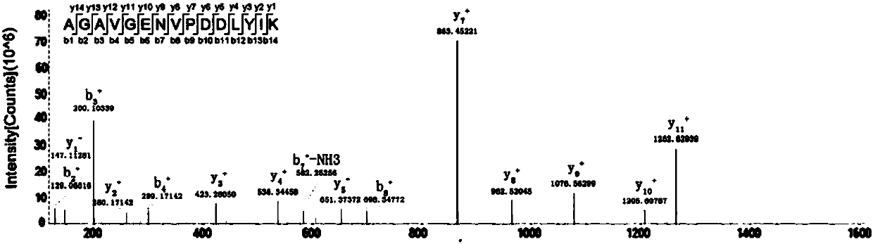
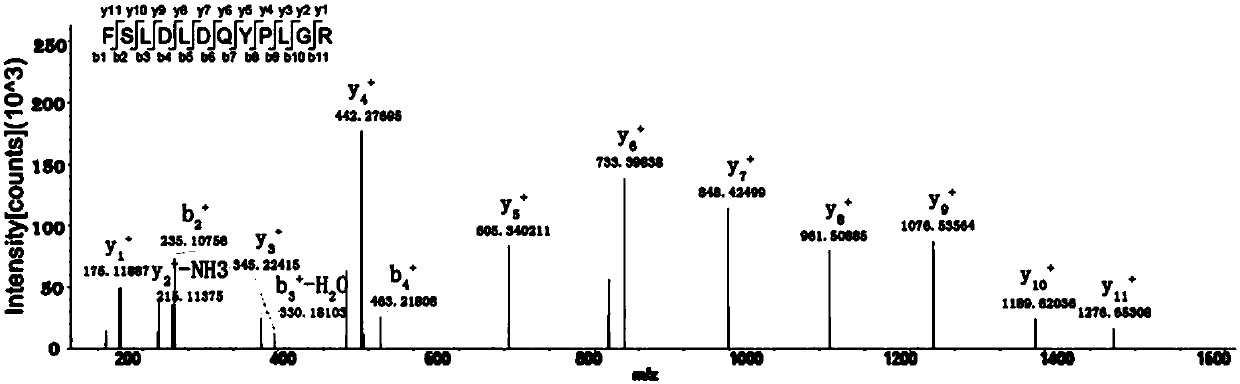
[0146] Example 1. Identification of HPV16 L1 and HPV18 L1 characteristic peptides
[0147] 1.1 Determination of candidate characteristic peptides
[0148] The inventors first selected trypsin that specifically cleaves the C-terminus of lysine and arginine, and used the PeptideMass module of ExPASy software to simulate the enzymes of HPV16 L1 and HPV18 L1 protein sequences (provided by Jiangsu Ruike Biotechnology Co., Ltd.) Then use the ClustalW module of Bioedit software to combine the above-mentioned simulated enzymolysis fragments with the L1 proteins involved in the types of HPV preventive vaccines (including HPV6, HPV11, HPV31, HPV33, HPV35, HPV39, HPV45, HPV51, HPV52, HPV56, HPV58, HPV59) were compared to obtain peptides (ie, candidate type-specific signal peptides / characteristic peptides) that only exist in theoretical hydrolysis products of HPV16 or HPV18 L1 proteins. The results are shown in Table 2 and Table 3 respectively. In theory, HPV16 L1 has 21 candidate type-s...
Embodiment 2
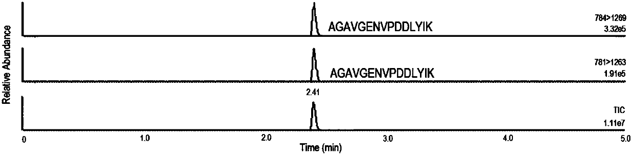
[0160] Example 2. Mass Spectrometry Quantitative Detection Method for Single Type HPV L1
[0161] In this example, the content of the HPV16 characteristic peptide and HPV18 characteristic peptide obtained in Example 1 was measured by isotope dilution method, so as to realize the quantification of HPV16 L1 protein and HPV18 L1 protein.
[0162] 2.1 Characteristic peptides and internal standard peptides
[0163] The sequences of the characteristic peptides of HPV16 L1 protein and HPV18 L1 protein and the corresponding isotope-labeled peptides (internal standard peptides) are shown in Table 4. The above polypeptides were all synthesized by China GL Biochem LTD and packaged into 1 mg / bottle.
[0164] Table 4: Type-specific peptides and internal standard peptides of HPV16 L1 and HPV18 L1
[0165]
[0166] 2.2 Standard curve
[0167] A mixed solution of HPV16 characteristic peptide and HPV18 characteristic peptide was prepared as an external standard, the concentration of both...
Embodiment 3
[0176] Example 3. Determination of HPV16 L1 and HPV18 L1 protein content in commercial products
[0177] The inventors used the isotope dilution mass spectrometry (abbreviated as IDMS) described in Example 2 to analyze four parts of HPV16 L1 stock solution before monovalent adsorption (Y1-16, Y2-16, E1-16 and I1-16), four parts of HPV18 L1 Stock solution before monovalent adsorption (Y1-18, Y2-18, E1-18 and I1-18), a part of HPV16 L1 stock solution after monovalent adsorption (E1-16-Al), and commercial vaccine Cervarix (purchased from GlaxoSmithKline (GSK)), Gardasil (Gardasil) and Gardasil 9 (Gardasil 9) (purchased from Merck & Co) in the HPV16 L1 and HPV18 L1 protein content were detected; and, while using The conventional BCA method and Bradford method in the field were used to measure the protein content of the above samples. The detection results of HPV16 L1 protein content are as follows: image 3 , Table 6, the detection results of HPV18 L1 protein content are as foll...
PUM
 Abstract
Description
Claims
Application Information
 - R&D
- Intellectual Property
- Life Sciences
- Materials
- Tech Scout
- Unparalleled Data Quality
- Higher Quality Content
- 60% Fewer Hallucinations
Browse by: Latest US Patents, China's latest patents, Technical Efficacy Thesaurus, Application Domain, Technology Topic, Popular Technical Reports.
© 2025 PatSnap. All rights reserved.Legal|Privacy policy|Modern Slavery Act Transparency Statement|Sitemap|About US| Contact US: help@patsnap.com