Method for coaxially preparing injectable PLGA drug-carrier microsphere by utilizing electrospinning machine

A technology of electrospinning machine and drug-loaded microspheres, which is applied in the direction of pharmaceutical formulations, medical preparations with non-active ingredients, medical preparations containing active ingredients, etc., can solve the problems of large toxic and side effects, and achieve biocompatibility Good, great research value and development prospect, realize the effect of slow and long-term release

Inactive Publication Date: 2018-05-29
JILIN UNIV
View PDF0 Cites 5 Cited by
  • Summary
  • Abstract
  • Description
  • Claims
  • Application Information

AI Technical Summary

Problems solved by technology

Although paclitaxel and etoposide have good anti-tumor effects, they have large toxic and side effects, mainly myelosuppression, cardiotoxicity and neurotoxicity. Now the research progress is in the direction of liposomes, which are used to reduce the neurotoxicity of anti-tumor drugs

Method used


Image

  • Method for coaxially preparing injectable PLGA drug-carrier microsphere by utilizing electrospinning machine
  • Method for coaxially preparing injectable PLGA drug-carrier microsphere by utilizing electrospinning machine
  • Method for coaxially preparing injectable PLGA drug-carrier microsphere by utilizing electrospinning machine

Examples


Embodiment 1

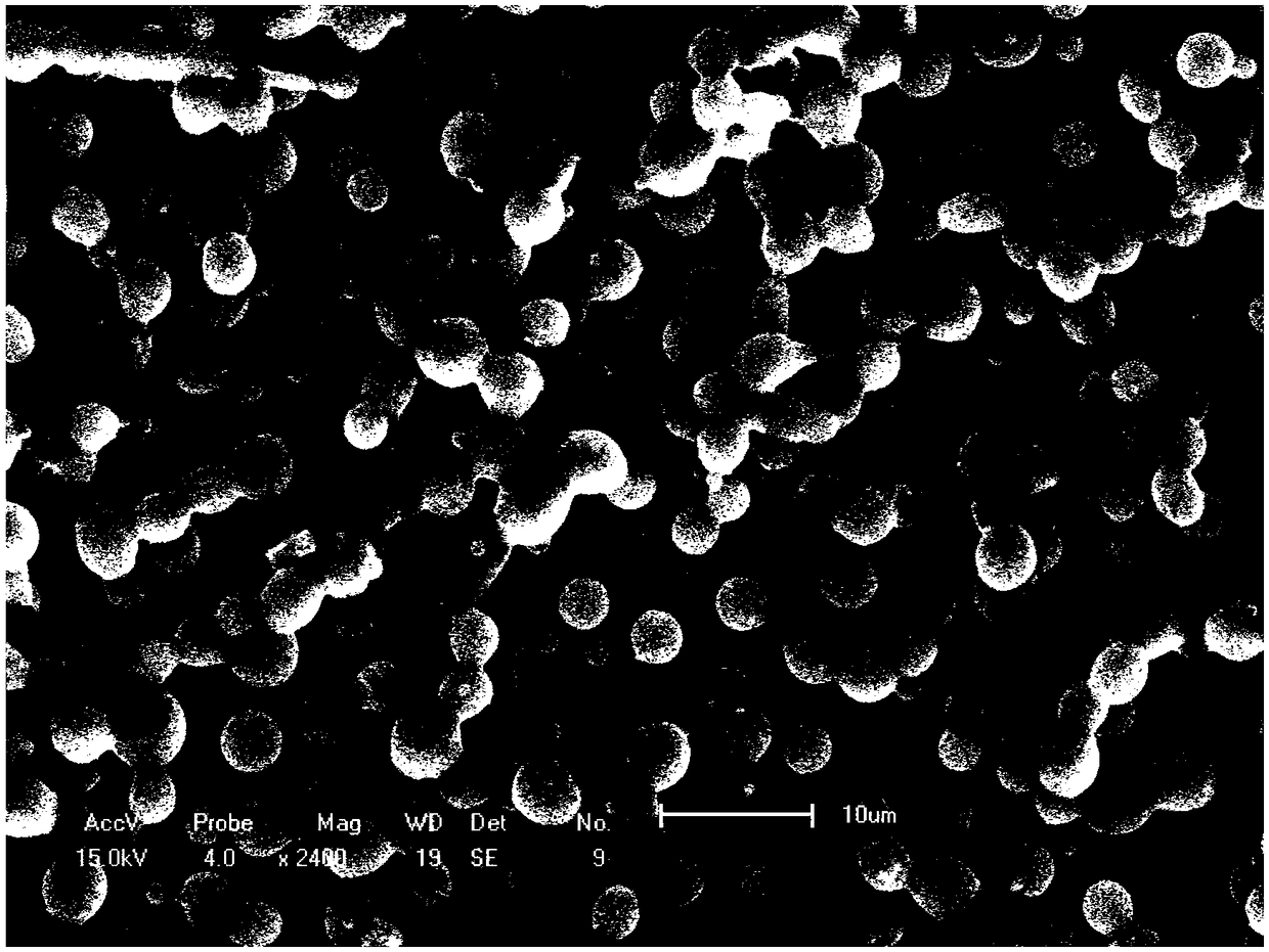
[0028] ① Weigh 60mg PLGA and dissolve it in 1ml hexafluoroisopropanol to prepare a 6% (g / ml) shell layer PLGA solution, stir magnetically for 4 hours at room temperature; prepare a certain concentration of PTX and ETP solutions with dichloromethane solution as Nuclei layer solution, vortexed to mix.

[0029] 2. Adjust the spinning voltage of electrospinning to be 10KV, the spinning distance to be 20cm, and the spinning speed shell layer and core layer to be 1.075ml / h and 0.12ml / h respectively, and then freeze-dry to obtain PLGA drug-loaded microspheres. Store at -20°C for later use. The scanning electron microscope results are as follows figure 1 As shown, the average particle size ranges from 1 to 4 μm, and the shape of the microspheres is relatively regular and there is adhesion.

Embodiment 2

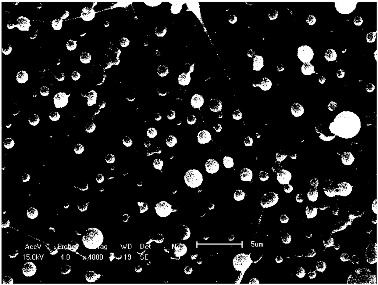
[0031] ①Weigh 50mg of PLGA and dissolve it in 1ml of hexafluoroisopropanol to prepare a 5% (g / ml) PLGA solution, stir magnetically at room temperature for 4 hours to obtain a uniform and stable shell solution; use dichloromethane solution to prepare a certain concentration The PTX and ETP solutions were vortexed to obtain the core layer solution.

[0032] ② Adjust the spinning voltage of the electrospinning machine to 15KV, the spinning distance to 15cm, and the spinning speed to 1.075ml / h and 0.12ml / h for the shell layer and the core layer, respectively, and obtain PLGA drug-loaded microspheres by high-voltage electrospinning, and freeze Store at -20°C after drying. The electron microscope results are as figure 2 As shown, the particle size ranges from 1 to 3.5 μm, the size is relatively uneven, and there are filaments.

Embodiment 3

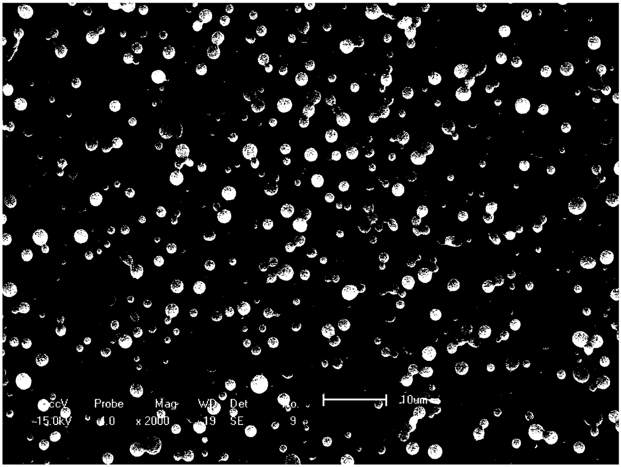
[0034] ①Weigh 50mg of PLGA and dissolve in 1ml of hexafluoroisopropanol solution to prepare a 5% (g / ml) PLGA solution, and stir magnetically for 4 hours at room temperature to obtain a uniform and stable shell solution; Concentration of PTX and ETP solution, after vortex mixing to obtain the core layer solution.

[0035] ② Adjust the spinning voltage of the electrospinning machine to 18KV, the spinning distance to 15cm, and the spinning speed to 1.075ml / h and 0.12ml / h for the shell layer and the core layer, respectively, and obtain PLGA drug-loaded microspheres by high-voltage electrospinning, and freeze Store at -20°C after drying. The electron microscope results are as image 3 As shown, the shape is relatively regular.


PUM

PropertyMeasurementUnit
particle diameteraaaaaaaaaa
particle sizeaaaaaaaaaa

Abstract

The invention belongs to preparation of drug-carrier microspheres, and discloses a method for coaxially preparing an injectable PLGA (poly(lactic-co-glycolic acid)) drug-carrier microsphere by utilizing an electrospinning machine. The method comprises the following steps: dissolving PLGA in hexafluoro isopropyl alcohol to prepare a PLGA solution as a shell layer solution; dissolving PTX (Paclitaxel) and ETP (Etoposide) in a dichloromethane solution to prepare a solution as a nuclear layer solution; stirring the two solutions and respectively injecting the solutions into a disposable 1ml plastic syringe; performing high-pressure electrostatic spraying by utilizing the electrospinning machine; then performing lyophilization to prepare PLGA microspheres with embedded PTX and ETP. The coaxialdrug-carrier microspheres prepared by the invention can be subjected to sustained-release and controlled-release and can be used for injecting. According to the method disclosed by the invention, through characteristic observation on the microspheres and determination on particle sizes of the microspheres, the average particle size of the drug-carrier microspheres is 3 microns, and the surfaces ofthe drug-carrier microspheres are smooth without holes. The composite microspheres prepared by the method disclosed by the invention are safe and free of toxicity, and can be used for postoperative treatment of osteosarcoma; a specific sustained-release effect is beneficial for reducing the damage of anti-tumor drugs to a human body, the anti-tumor activity of the drugs is improved, and the microspheres have a broad research value and development prospects.

Description

technical field [0001] The invention belongs to biological tissue engineering cell scaffold materials, in particular to a preparation method of PLGA drug-loaded microspheres with an anti-osteosarcoma effect, in particular to a method for preparing coaxial PLGA drug-loaded microspheres with slow-release effect through an electrospinning machine method. Background technique [0002] Osteosarcoma (OS) is a malignant connective tissue tumor, and the incidence of osteosarcoma occupies the first place among malignant tumors. Osteosarcoma is a common, highly malignant and poor prognosis of malignant bone tumors. After a few months, it can spread to the lungs. Three years after amputation, the survival rate is only 5% to 20%. Therefore, we should speak highly of it. So far, many authors have used cisplatin (DDP), doxorubicin (ADR), methionine folate (mtx-cf), BCD, ifosfamide (IFO) and cyclophosphamide ( CTX) etc. to treat osteosarcoma, and the disease-free survival rate is 42%....

Claims


Application Information

Patent Timeline
Patent Type & Authority Applications(China)
IPC IPC(8): A61K9/50A61K47/34A61K31/337A61K31/7048A61P35/00
CPCA61K9/5031A61K9/0024A61K31/337A61K31/7048A61K2300/00
Inventor 张梅唐亚军朱振华赵贺姚霁航
Owner JILIN UNIV
Why Eureka
Try Eureka