Fast separation method for placenta hematopoietic stem cells

A technology of hematopoietic stem cells and separation methods, applied in the field of rapid separation of placental hematopoietic stem cells, can solve the problems of perfusate perfusion difficulties, formation of fibrosis, long time, etc., achieve the effect of enriching the source of stem cells, avoiding the formation of fibrosis, and simple operation

Inactive Publication Date: 2018-06-12
ACADEMY OF MILITARY MEDICAL SCI +1
View PDF6 Cites 4 Cited by
  • Summary
  • Abstract
  • Description
  • Claims
  • Application Information

AI Technical Summary

Problems solved by technology

This preparation method mainly adopts the method of perfusion from placental umbilical vein and artery, which has disadvantages such as long time, difficulty in accurately finding umbilical artery and umbilical vein, difficulty in perfusate perfusion, and easy blood coagulation in actual ope...

Method used


Image

  • Fast separation method for placenta hematopoietic stem cells
  • Fast separation method for placenta hematopoietic stem cells
  • Fast separation method for placenta hematopoietic stem cells

Examples


Embodiment 1

[0031] Example 1: Isolation method of placental HSC

[0032] Take 450-600g placenta of normal delivery under sterile conditions within 4-6 hours after delivery, soak it in PBS buffer, store it at room temperature for 2-6 hours and process it as soon as possible; remove the placenta from the soaked PBS buffer in a biological safety cabinet Take out the solution, place it in another sterile surgical tray, add 800-1000mL of fresh PBS buffer solution to soak it completely, and use surgical scissors to completely cut the placenta leaflet, wash the placenta leaflet fully with PBS buffer solution, and wash out the residual placental leaflet blood, and quickly collect the flushing liquid; filter the flushing liquid with a 200-mesh copper mesh, collect the filtered cell suspension, and add it to multiple 50ml centrifuge tubes, centrifuge at 900g for 10 minutes, pour off the supernatant, and use 10% Volume fetal bovine serum (FBS) in PBS buffer to resuspend the cells; then add the cell ...

Embodiment 2

[0034] Example 2: Cryopreservation of placental HSC

[0035] After counting the isolated and purified hematopoietic stem cells with trypan blue, the 6 The cells were added to 1 ml of cell freezing medium (containing 50% IMEM culture medium, 40% FBS, and 10% dimethyl sulfoxide), and after programmed cooling, they were finally put into liquid nitrogen tubes for freezing.

Embodiment 3

[0036] Example 3: Biological Characterization of Placental HSCs

[0037] 1. Cell Morphological Characteristics

[0038] Through the separation and purification of Example 1, the placental mononuclear cells can be seen as round suspension cells under a microscope ( figure 1 -A), using an inverted high-power microscope to observe hematopoietic stem cells, it can be seen that the cells grow in a typical round suspension ( figure 1 -B).

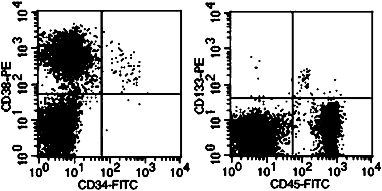
[0039] 2. Identification of placental HSC surface markers by flow cytometry


PUM


Abstract

The invention discloses a fast separation method for placenta hematopoietic stem cells. The method specifically includes: completely soaking the placenta obtained after normal labor into a buffer solution; cutting the placenta lobule into pieces, and using a buffer solution to sufficiently wash the placenta lobule, and collecting the washing liquid; filtering with a copper screen, and collecting filtrate; using a density gradient centrifugation method to centrifugally separate mononuclear cells, washing with a buffer solution, and using a hematopoietic stem cell culture medium to suspend the acquired cells to obtain the placenta hematopoietic stem cells. The placenta hematopoietic stem cells have the same biological function as umbilical cord blood-source hematopoietic stem cells, and thenumber of the acquired hematopoietic stem cells can satisfy the needs of adult transplant. The separation method has the advantages that the method is simple to operate, convenient and practical; theplacenta hematopoietic stem cells are young in components, wide in source, convenient and easy to obtain and promising in application prospect in stem cell clinical application.

Description

technical field [0001] The invention belongs to the technical field of hematopoietic stem cell separation, and in particular relates to a rapid separation method of placental hematopoietic stem cells. Background technique [0002] Hematopoietic stem cells (HSCs) were first isolated from bone marrow. They are a type of adult stem cells derived from mesoderm with multi-lineage differentiation potential and self-renewal ability. Differentiation ability of cells of various lineages, mainly including myeloid, lymphoid, megakaryotic and so on. The latest research shows that hematopoietic stem cells not only play an important role in rebuilding the body's hematopoietic system and immune system, but also use genetic engineering technology to introduce foreign genes to treat related diseases and have achieved remarkable curative effects. Therefore, hematopoietic stem cells have broad application prospects in future clinical treatment. [0003] Currently reported hematopoietic stem ...

Claims


Application Information

Patent Timeline
IPC IPC(8): C12N5/0789C12N5/073
CPCC12N5/0605C12N5/0647C12N2509/00
Inventor 张毅刘元林刘伟江李雪杨焕凤
Owner ACADEMY OF MILITARY MEDICAL SCI
Why Eureka
Try Eureka