Method for rapid multi-nozzle 3D printing of tumor tissue model using supporting bath

A tumor tissue, 3D printing technology, applied in the field of biological 3D printing, can solve the problems of increasing the bioprinting time, restoring the tumor tissue structure, accuracy and time effects, avoiding the uncontrollable cell density and species space, and slowing down the activity. The effect of reducing, reducing printing time

Active Publication Date: 2018-09-28
JINAN UNIVERSITY
View PDF3 Cites 15 Cited by
  • Summary
  • Abstract
  • Description
  • Claims
  • Application Information

AI Technical Summary

Problems solved by technology

(Ratna and Karger-Kocsis 2008, Du 2015) patent 201610830529.5 uses hyaluronic acid, sodium alginate, GelMA (light-cured gelatin), transglutaminase and brain tumor cells to directly mix 3D printing and in vitro culture of brain tumor in vitro models, However, with the increase in the number of printing layers, the support strength is insufficient, and the tumor tissue structure cannot be well restored.
And this study only prints tumor cells, and does not show normal tissue cells, and cannot completely restore the structure of tumor tissue
Common 3D bioprinting methods cannot satisfy the sterile environment of cells well, and the processing of spatial configuration and internal structure is not ideal
[0003] When printing a variety of cell materials, the nozzle replacement procedure is cumbersome, which increases the time of bioprinting and has a great impact on its accuracy and time.
What's more, it will cause the reduction of cell activity, which will bring huge resistance to multi-cellular bioprinting.

Method used


Image

  • Method for rapid multi-nozzle 3D printing of tumor tissue model using supporting bath

Examples


Embodiment 1

[0036] Preparation of Example 1 Liver Tumor Tissue Model

[0037] (1) Preparation of liver tumor cell ink

[0038] With 25mM HEPES buffer solution containing 10% volume FBS as solvent, configure GelMA (photocurable gelatin) 2% and sodium alginate 2% magnetically stir evenly, sterilize with ultraviolet light, add 0.1% Irgacure 2959 photoinitiator; Tumor cells (2×10 6 / mL) DMEM complete medium, pipet evenly with a pipette gun, and put it into the incubator for use.

[0039] (2) Preparation of liver tissue ink

[0040] With 25mM HEPES buffer solution containing 10% volume FBS as solvent, configure GelMA (photocurable gelatin) 2% and sodium alginate 2% magnetically stir evenly, sterilize with ultraviolet light, add 0.1% Irgacure 2959 photoinitiator; Tissue cells (2×10 6 / mL) into the DMEM complete medium, blow evenly with a pipette gun, and put it into the incubator for use.

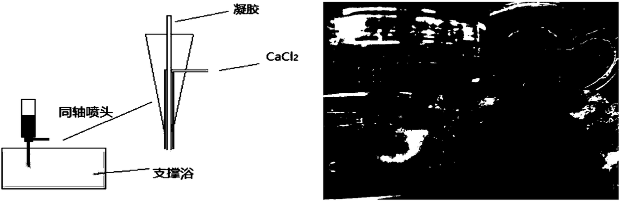
[0041] (3) Preparation of hydrogel support bath

[0042] The main material of the hydrogel support ...

Embodiment 2

[0045] The preparation of embodiment 2 lung tumor tissue model

[0046] (1) Preparation of lung tumor cell ink

[0047] With 25mM HEPES buffer solution containing 10% volume FBS as solvent, configure GelMA (photocurable gelatin) 3% and sodium alginate 2% magnetically stir evenly, sterilize with ultraviolet light, add 0.1% Irgacure 2959 photoinitiator; add lung Tumor cells (3×10 6 / mL) DMEM complete medium, pipet evenly with a pipette gun, and put it into the incubator for use.

[0048] (2) Preparation of lung tissue cell ink

[0049]With 25mM HEPES buffer solution containing 10% volume FBS as solvent, configure GelMA (photocurable gelatin) 3% and sodium alginate 2% magnetically stir evenly, sterilize with ultraviolet light, add 0.1% Irgacure 2959 photoinitiator; lung normal Tissue cells (3×10 6 / mL) into the DMEM complete medium, blow evenly with a pipette gun, and put it into the incubator for use.

[0050] (3) Preparation of hydrogel support bath

[0051] The main mate...

Embodiment 3

[0054] Example 3 Preparation of Pancreatic Tumor Tissue Model

[0055] (1) Preparation of pancreatic tumor cell ink

[0056] With 25mM HEPES buffer solution containing 10% volume FBS as solvent, configure GelMA (photocurable gelatin) 4% and sodium alginate 1% magnetically stir evenly, sterilize with ultraviolet light, add 0.1% Irgacure 2959 photoinitiator; add pancreas containing Tumor cells (2×10 6 / mL) DMEM complete medium, pipet evenly with a pipette gun, and put it into the incubator for use.

[0057] (2) Preparation of pancreatic tissue cell ink

[0058] With 25mM HEPES buffer solution containing 10% volume FBS as solvent, configure GelMA (photocurable gelatin) 4% and sodium alginate 1% magnetically stir evenly, sterilize with ultraviolet light, add 0.1% Irgacure 2959 photoinitiator; normal pancreas Tissue cells (2×10 6 / mL) into the DMEM complete medium, blow evenly with a pipette gun, and put it into the incubator for use.

[0059] (3) Preparation of hydrogel suppo...


PUM


Abstract

The invention discloses a method for rapid multi-nozzle 3D printing of a tumor tissue model using supporting bath, and relates to the field of biological 3D printing. The method mixes alginate, photocurable gelatin and the like to encapsulate cells and prints rapidly in the supporting bath, and printing of different cells is achieved by rapidly switching nozzles. By printing a normal tissue and atumor tissue in a complete model, the structure of a tumor tissue in vivo is better restored. The closed property of the hydrogel support bath provides a better sterile environment, and at the same time, uncontrolled cell density and species space due to settlement are avoided during printing. The rapid switching of the nozzles reduces the printing time to a certain extent, the decreasing of the activity of the cells is effectively slowed down in the printing process, and the integrity of the printing structure is effectively ensured. The problem of gel curing is effectively solved by printingwith a concentric needle. The tumor model is built faster and can be better used for tumor treatment research.

Description

technical field [0001] The invention relates to the field of biological 3D printing, in particular to a method for rapidly 3D printing a tumor tissue model using a multi-nozzle support bath. Background technique [0002] Tumor tissue models generally need to be constructed by planting tumor cells in organisms such as mice, which greatly wastes manpower and material resources. With the development of bio-3D printing technology, its application in cell culture, tissue engineering and other fields is increasing, but its application in tumor models is still lack of research. Compared with tumor models constructed by traditional methods, the three-dimensional structure of tumors constructed by 3D printing can better reflect the growth and development of tumors in vivo, and it is a tumor model that is closer to the pathological characteristics of cancer cells in vivo. Du, Xinru and others used 3D printing technology to create a drug screening model, providing a certain technical ...

Claims


Application Information

Patent Timeline
Patent Type & Authority Applications(China)
IPC IPC(8): C12M3/00B33Y30/00
CPCB33Y30/00C12M33/00
Inventor 薛巍宋镕光阮淼亮戴箭王永周
Owner JINAN UNIVERSITY
Why Eureka
Try Eureka