
Amplification method of umbilical cord mesenchymal stem cells and application of umbilical cord mesenchymal stem cells in arthritis

A technology of mesenchymal stem cells and umbilical cords, applied in the field of osteoarthritis treatment, can solve the problems of limited expansion capacity and achieve the effect of fast expansion

Active Publication Date: 2018-09-28
杨凌洛威塔生物科技有限责任公司
View PDF2 Cites 4 Cited by
  • Summary
  • Abstract
  • Description
  • Claims
  • Application Information

AI Technical Summary

Problems solved by technology

[0006] The purpose of the present invention is to provide a method for the expansion of umbilical cord mesenchymal stem cells to solve the problems of limited expansion ability in the cultivation of umbilical cord mesenchymal stem cells in the actual application process, and to use the mesenchymal stem cells combined with other components Joint treatment of osteoarthritis for better treatment results

Method used


Image

  • Amplification method of umbilical cord mesenchymal stem cells and application of umbilical cord mesenchymal stem cells in arthritis
  • Amplification method of umbilical cord mesenchymal stem cells and application of umbilical cord mesenchymal stem cells in arthritis
  • Amplification method of umbilical cord mesenchymal stem cells and application of umbilical cord mesenchymal stem cells in arthritis

Examples


Embodiment 1

[0040] Example 1: Isolation and culture of mesenchymal stem cells. In the operating room, the umbilical cord is removed, stored in sterile saline, and transported to the laboratory on a cold chain. On the 100-level ultra-clean workbench of the GMP laboratory, use sterile surgical instruments to peel off Wharton's jelly, add it to 10 times the volume of trypsin digestion solution (0.5% trypsin + 0.2% EDTA), and keep at 37 degrees Lower digestion 2h. Then add 1mg / mL type I collagen and continue to digest for 1h. Pass the above suspension through a 200-mesh sieve, blow and filter. The above cell suspension was mixed with the culture medium, resuspended, added to a T25 cell culture flask, and cultured in a 37°C, 5% carbon dioxide incubator until generation P1.

Embodiment 2

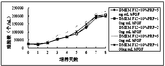
[0041] Example 2: Divide the experiment into five groups, Group A (DMEM / F12+ 10%PRP+5ng / mL bFGF), Group B (DMEM / F12+ 10%PRP+10ng / mL BFGF), Group C (DMEM / F12+ 10 %PRP + 20ng / mL bFGF), group D (DMEM / F12+10%PRP + 50ng / mL bFGF), group E (DMEM / F12+ 10%PRP + 100ng / mL bFGF).

[0042] After centrifugation, the P1 generation cells were resuspended in DMEM / F12 medium containing 10% PRP, and the density was adjusted to 5×10 4 cells / mL, inoculate five 24-well plates at 500 μL / well. According to the grouping, in the corresponding wells, add different concentrations of bFGF, 24 wells in each group. Subsequently, five 24-well plates were placed in a 37-degree, 5% carbon dioxide incubator for cultivation, and were cultured at 1 d, 2 d, 3 d, 4 d, 5 d, 6 d, 7 d, 8 d, a total of 8 time points , cells from 3 wells in each group were taken at each time point for cell counting.

[0043] figure 1 The experimental results showed that the cells in group D proliferated the fastest. It shows that t...

Embodiment 3

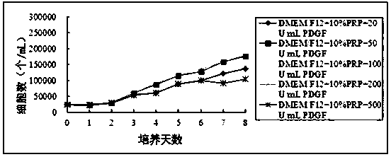
[0044] Example 3: Divide the experiment into five groups, group A (DMEM / F12+ 10%PRP + 20U / mL PDGF), group B (DMEM / F12+ 10%PRP + 50U / mL PDGF), group C (DMEM / F12+ 10 %PRP + 100U / mL PDGF), group D (DMEM / F12+10%PRP + 200U / mL PDGF), group E (DMEM / F12+ 10%PRP + 500U / mL PDGF).

[0045] After centrifugation, the P1 generation cells were resuspended in DMEM / F12 medium containing 10% PRP, and the density was adjusted to 5×10 4 cells / mL, inoculate five 24-well plates at 500 μL / well. According to the grouping, different concentrations of PDGF were added to the corresponding wells, 24 wells in each group. Subsequently, five 24-well plates were placed in a 37-degree, 5% carbon dioxide incubator for cultivation, and were cultured at 1 d, 2 d, 3 d, 4 d, 5 d, 6 d, 7 d, 8 d, a total of 8 time points , cells from 3 wells in each group were taken at each time point for cell counting.

[0046] figure 2 The experimental results showed that the cell proliferation in group C was the fastest. It...


PUM


Abstract

The invention relates to improvement of an isolated culture technology of umbilical cord mesenchymal stem cells and an application of the umbilical cord mesenchymal stem cells in arthritis. The isolated culture method of the umbilical cord derived mesenchymal stem cells and the application of the umbilical cord mesenchymal stem cells in the arthritis comprise the following steps: 1) obtaining themesenchymal stem cells from a human umbilical cord via an enzyme digestion method; 2) implementing culture of the mesenchymal stem cells via a DMEM / F12 medium which is added with basic fibroblast growth factors and platelet-derived growth factors in the combination with blood platelet enriched plasma; 3) improvement of hyaluronic acid: adding methacrylic anhydride to the hyaluronic acid, so that a3D bio-stent is formed, and subsequently, the mesenchymal stem cells can be immobilized to an osteoarticular injury site for conducting fixed-point repair; and 4) injecting the cultivated mesenchymalstem cells, together with PRP and the improved hyaluronic acid, into a bone joint of a volunteer, so as to treat osteoarthritis.

Description

technical field [0001] The invention relates to a method for expanding umbilical cord mesenchymal stem cells, and using the mesenchymal stem cells in combination with other components to treat osteoarthritis. Background technique [0002] Osteoarthritis is a progressive and painful disease that can affect both the young and the elderly. In an aging society, the prevalence is likely to increase further. It is estimated that at least 100 million people nationwide are affected by arthritis. Worldwide, arthritis is the fourth leading cause of disability. Osteoarthritis is an important cause of disability in both developed and developing countries. [0003] Osteoarthritis is a progressive and irreversible degenerative disease of cartilage. Articular cartilage is very poorly repairable and avascular, so lack of systemic regulation may lead to ineffective healing. There is evidence that osteoarthritis is associated with depletion of stromal mesenchymal stem cells, manifested b...

Claims


Application Information

Patent Timeline
Patent Type & Authority Applications(China)
IPC IPC(8): C12N5/0775A61K35/28A61K35/16A61K47/36A61P19/02A61P29/00C08B37/08
CPCA61K9/0019A61K35/16A61K35/28A61K47/36A61P19/02A61P29/00C08B37/0072C12N5/0668C12N2501/115C12N2501/135C12N2509/00A61K2300/00
Inventor 刘建强何晓艾霞
Owner 杨凌洛威塔生物科技有限责任公司
Learn More