Systems and methods for producing gastrointestinal tissues

A main body and cell technology, applied in biochemical equipment and methods, methods of supporting/immobilizing microorganisms, tissue regeneration, etc., can solve problems such as high morbidity and mortality

Active Publication Date: 2018-09-28
BIOSTAGE INC
View PDF7 Cites 4 Cited by
  • Summary
  • Abstract
  • Description
  • Claims
  • Application Information

AI Technical Summary

Problems solved by technology

But these treatments have high morbidity and mortality

Method used


Image

  • Systems and methods for producing gastrointestinal tissues
  • Systems and methods for producing gastrointestinal tissues
  • Systems and methods for producing gastrointestinal tissues

Examples


Embodiment

[0110] Example I: Esophageal Stent

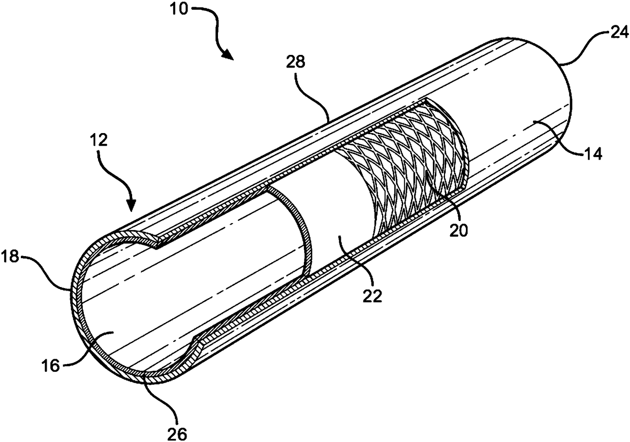
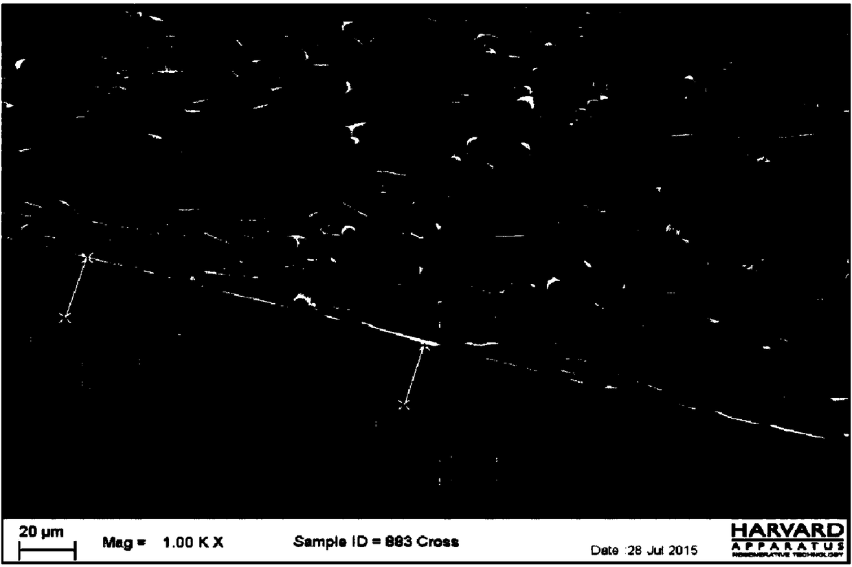
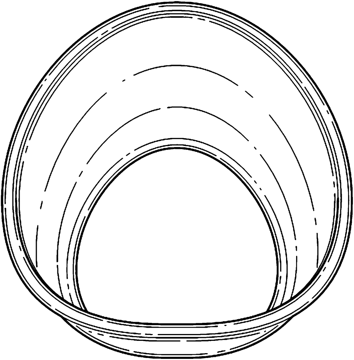
[0111] like Figure 1A As shown, a synthetic esophageal stent comprising three layers of material was prepared. A first layer of polyurethane (PU) is deposited by electrostatic atomization onto a metal mandrel, and then a braided material is deposited on top of the first PU layer. The second PU layer is then deposited by electrospinning. The formed scaffold is then removed from the mandrel. Each scaffold defines a tubular structure having three layers (inner electrostatically atomized layer, outer electrospun layer, and braid layer sandwiched between the inner electrostatically atomized layer and outer electrospun layer) the wall. The physical dimensions of the scaffolds were determined by scanning electron microscopy (SEM). The average scaffold wall thickness is about 500 microns. Figure 1B A non-limiting SEM view of a cross-section of the wall is shown in . Figure 1C A non-limiting view of a cross-section of a tubular stent is sh...

Embodiment II

[0120] Example II: Esophageal Implantation

[0121] like Figure 1A As shown, a synthetic esophageal stent comprising a three-layer material with an outer electrospun layer of polycarbonate-polyurethane prepared by dissolving in hexafluoroisopropanol (HFIP, DuPont, Wilmington , Delaware, USA) in 12% w / v polycarbonate polyurethane solution deposited. The electrospinning device used was purchased from IME Technologies, Geldrop (Netherlands), the Netherlands. Electrospun fibers were collected on a target aluminum mandrel rotating at 800 rpm and placed 22 mm from the syringe tip to deposit isotropic fibers, resulting in scaffolds with an average wall thickness of 500 μm. The scaffolds were dried in vacuum Removal of residual solvent. Then by a low-pressure plasma system (Diener Tetra 150-LF-PC-D), plasma treatment is carried out to the support with ethylene and oxygen gas of 2 forward cycles. The support is carried out gamma sterilization (Think Teri Corporation (STERIS), Northb...

Embodiment III

[0147] Example III - Other gastrointestinal implants

[0148] The procedures described in Example I and Example II may be performed instead in regions of the gastrointestinal tract confined to the rectum. The results were similar to those outlined previously.


PUM

PropertyMeasurementUnit
diameteraaaaaaaaaa
diameteraaaaaaaaaa
lengthaaaaaaaaaa

Abstract

Aspects of the disclosure relate methods and synthetic scaffolds for regenerating gastrointestinal tissue (e.g., esophageal tissue).

Description

[0001] References to related applications [0002] This application claims priority to U.S. Provisional Patent Application No. 62 / 254,700 filed on November 12, 2015 and U.S. Provisional Patent Application No. 62 / 276,715 filed on January 8, 2016 , the entire disclosure of the aforementioned prior application is hereby incorporated by reference into this application. technical field [0003] The present invention relates to engineered tissue for replacing or repairing damaged tissue. Background technique [0004] Engineered biological tissues for the replacement or repair of damaged tissue are typically produced by seeding cells on synthetic scaffolds and exposing the cells to conditions that allow them to synthesize and secrete extracellular matrix components on the scaffold. Different techniques have been used to generate synthetic scaffolds, including nanofiber assembly, casting, printing, physical jetting (e.g., using pumps and syringes), electrospinning, electrostatic a...

Claims


Application Information

Patent Timeline
Patent Type & Authority Applications(China)
IPC IPC(8): A61F2/04A61L27/14A61L27/56
CPCA61F2/04A61F2/0077A61L27/3882A61L27/3895A61L27/3834A61L27/18A61L27/04A61L27/56A61L27/58C12N5/0662C12N5/0653A61F2002/044A61F2002/045A61F2002/043A61F2002/046A61L2430/22C12N2535/00C12N2533/30C08L75/04C08L67/02A61F2/90C12M21/08C12M25/14A61F2002/0086A61F2210/0004A61F2210/0076A61F2230/0069A61B17/320016A61F2/07C12N5/0068C12N5/0697A61F2250/0067A61F2/82
Inventor 萨韦里奥·香格里拉弗朗西丝卡谢里夫·苏莱曼马修·马什
Owner BIOSTAGE INC
Why Eureka
Try Eureka