Cell telescoping structure model induced through mitosis retarding and preparation method thereof

A cell model, mitotic technology, applied in the fields of biochemical equipment and methods, analytical materials, and microbial determination/inspection, which can solve problems such as rare, difficult to obtain cell-in-cell structure, etc.

Inactive Publication Date: 2018-10-30
ACADEMY OF MILITARY MEDICAL SCI
View PDF1 Cites 3 Cited by
  • Summary
  • Abstract
  • Description
  • Claims
  • Application Information

AI Technical Summary

Problems solved by technology

[0004] In addition, it is worth noting that the cell-in-cell phenomenon is rarely seen in norma

Method used


Image

  • Cell telescoping structure model induced through mitosis retarding and preparation method thereof
  • Cell telescoping structure model induced through mitosis retarding and preparation method thereof
  • Cell telescoping structure model induced through mitosis retarding and preparation method thereof

Examples


Embodiment 1

[0041] Embodiment 1, establishment of MCF10A / H2B-mCherry stable cell line

[0042] 1. Cell laying

[0043] Firstly, 293FT cells were mixed with 1×10 per well 6 The density of each cell was evenly spread on a six-well plate pre-embedded with collagen (collagen I), and the medium DMEM (containing 10% FBS, % indicates volume percentage) was filled to 2 mL, and cultured in an incubator for 16 hours. Transfection experiments;

[0044] 2. Retroviral packaging system

[0045] Solution A: Mix 0.4μg target plasmid pBabe-H2B-mCherry, 0.2μg pCMV-VSV-G plasmid, 0.25μg gag / pol plasmid with 100μL Opti-MEM, and let stand at room temperature for 5min; Lipofectamine 2000 was mixed by inverting gently. Dilute 2.5 μL Lipofectamine 2000 into 100 μL Opti-MEM, mix by inverting gently, and let it stand at room temperature for 5 minutes. Leave it for 20-30 minutes; mix 800 μL Opti-MEM with 200 μL solution C to obtain transfection mixture D.

[0046] 3. Packaging of retroviruses

[0047] Discard...

Embodiment 2

[0054] Embodiment 2, the statistical method of cell-in-cell formation rate

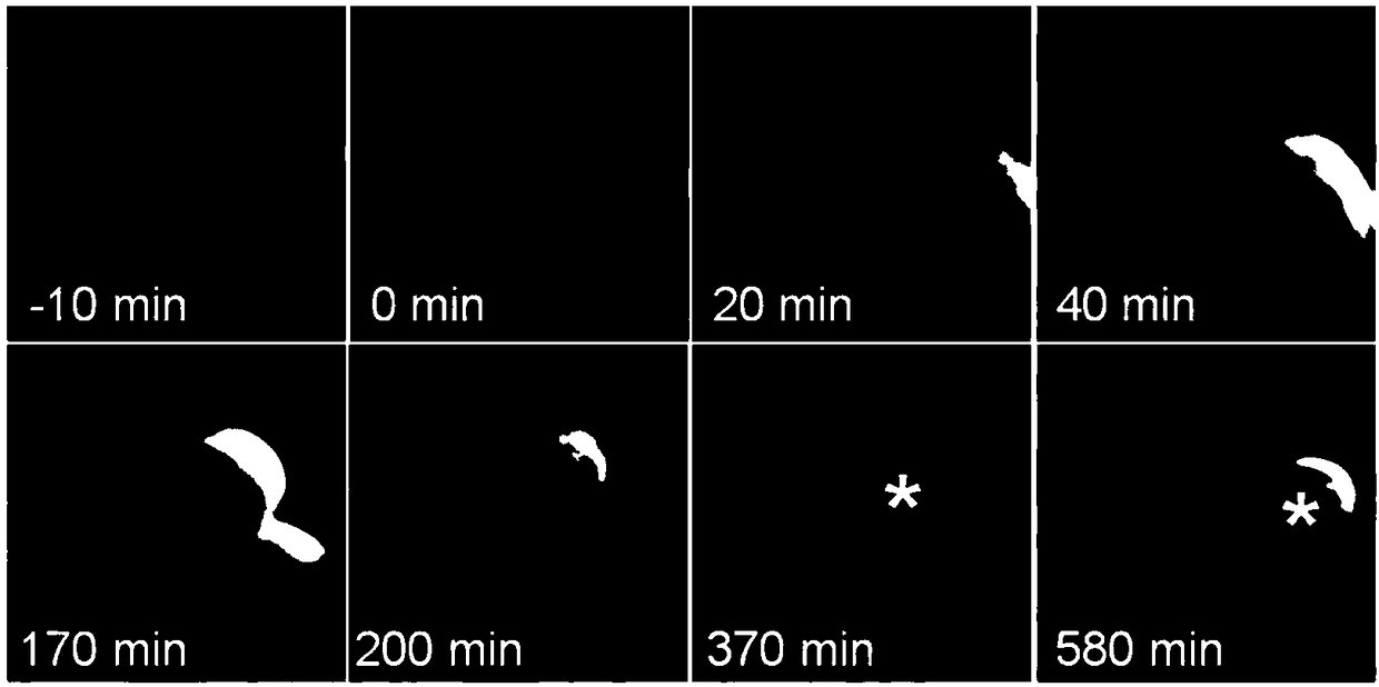
[0055] Cell-in-cell structure such as figure 1 As shown, the red fluorescent H2B-mCherry marks the nucleus, and the cell-in-cell formation process is tracked by the live cell workstation time-lapse, and the yellow * indicates the internal cells of the cell-in-cell structure. figure 1 Schematic diagram of Time-lapse for the formation of cell-in-cell structures induced by targeted arrest of mitotic metaphase. In a specific embodiment, the total number of cells in the shooting field of view is counted using the NIS-Elements system supporting the Nikon microscope, and the number of cells is identified according to the H2B-mCherry labeled nucleus; the structure of the internal cells completely wrapped by the target cells (ie external cells) is recorded as 1 cell-in-cell structure. cell-in-cell (%)=number of cell-in-cells / total number of cells×100 mean±SD.

Embodiment 3

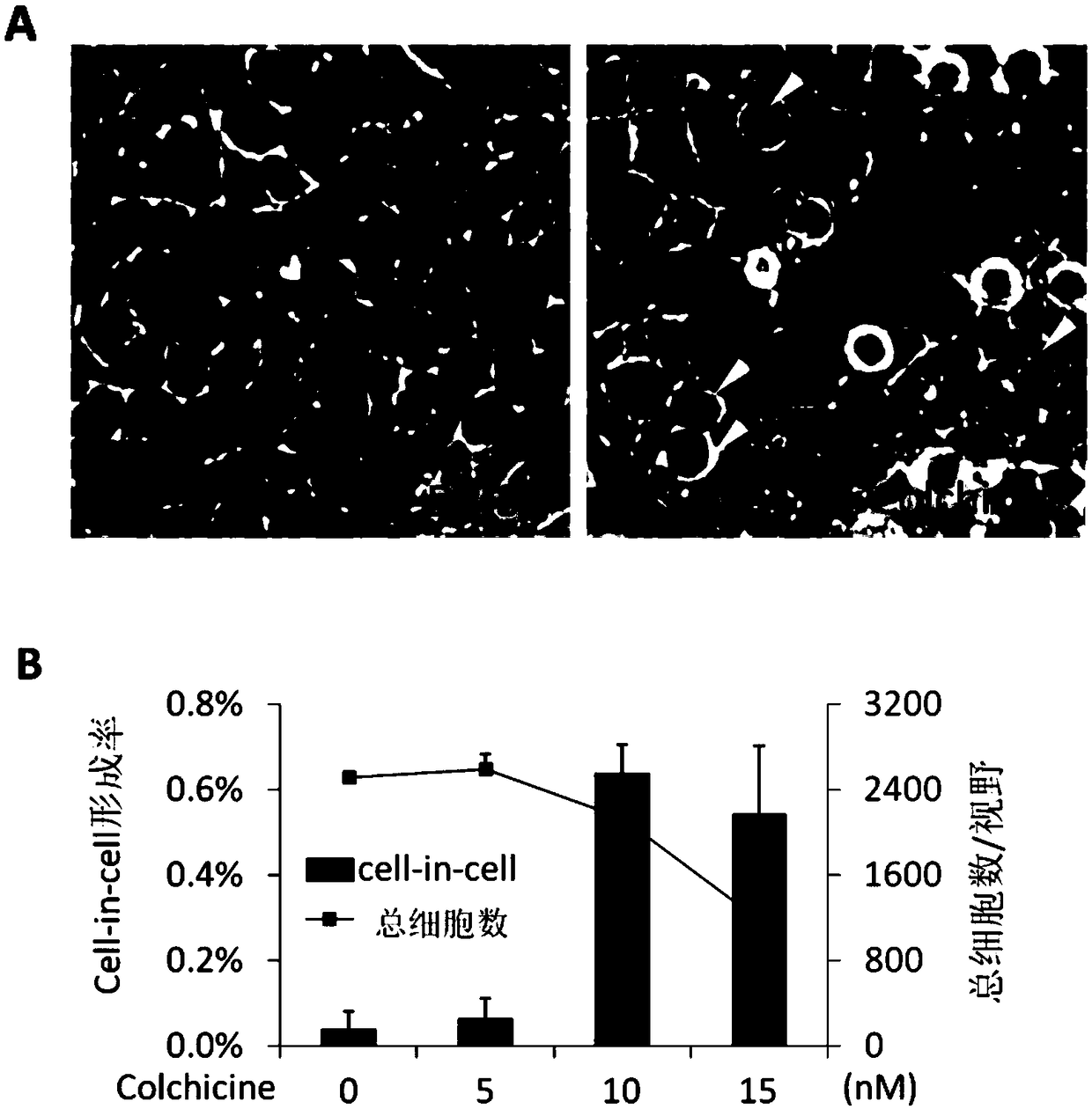
[0056] Example 3, colchicine induces cell-in-cell structure formation

[0057] Inoculate the stable cell line MCF10A / H2B-mCherry prepared in Example 1 with a good culture state and a confluence of more than 80% into a 12-well glass bottom cell culture plate at a cell density of 1×10 5 / hole. After the cells adhered to the wall, the medium containing colchicine, a mitosis inhibitor, was replaced, and the following concentration gradients of 0 nM, 5 nM, 10 nM and 15 nM were set. Colchicine was treated for 3 days, and the medium was changed every day. After 3 days of drug treatment, discard the medium carefully, add 1ml 1×PBS to wash twice, fix with 4% paraformaldehyde, discard the fixative after 10min, add PBS to wash twice, and finally add DAPI dropwise to seal the slide. Cell morphology and cell-in-cell formation were observed under a Nikon inverted fluorescence microscope, and photographed and recorded. Set up a 10× objective lens, two-channel shooting: mCherry and DIC, ra...


PUM


Abstract

The invention discloses a cell telescoping structure model induced through mitosis retarding and a preparation method thereof. The method includes: retarding the mitosis metaphase through chemical drug or a genetics method to obtain a CIC cell model. The CIC cell model is induced through targeted retarding of mitosis. Building of the cell model is not limited in treatment mode and can be realizedno matter through the chemical drug or through the genetics method, and a stable and reliable model is provided for studying formation mechanism and physiological and pathological significance of CIC.Mitosis retarding induced CIC structure can be realized in normal epithelial cells, and the CIC cell model can be built in tumor cells. The CIC cell model can be built by using tumor targeting drug for targeted retarding of the mitosis metaphase, it shows that the model can serve as an in-vitro model for tumor therapy research.

Description

technical field [0001] The invention belongs to the field of biotechnology, and relates to a cell nesting structure model induced by mitosis arrest and a preparation method thereof. Background technique [0002] Cell-in-cell structure (cell-in-cell structure or CIC structure) refers to a unique cell-in-cell-like structure formed by one or more living cells located inside another cell. Interaction is a complex process that produces biological effects. Studies on clinical tissue samples have shown that cell-in-cell structures are commonly found in human tumor tissues, such as breast cancer, colon cancer, cervical cancer, prostate cancer, liver cancer, etc., but are rarely seen in epithelial or normal tissues, suggesting that this phenomenon is similar to human tumors. tumors are closely related. After the homogeneous cell-in-cell structure is formed, the fate of inner and outer cells is diversified, but the most important thing is that inner cells die in their target cells (...

Claims


Application Information

Patent Timeline
IPC IPC(8): C12Q1/02
CPCG01N33/5008G01N33/5011
Inventor 孙强梁剑青牛祖彪于晓晨
Owner ACADEMY OF MILITARY MEDICAL SCI
Why Eureka
Try Eureka