
Test strip for identification and detection of bovine viral diarrhea virus and preparation method thereof

A technique for bovine viral diarrhea and test strips, which is applied to measurement devices, instruments, scientific instruments, etc., can solve the problems of limited application, complicated operation, high price, etc. Effect

Active Publication Date: 2021-03-12
河南中泽生物工程有限公司 +1
View PDF12 Cites 0 Cited by
  • Summary
  • Abstract
  • Description
  • Claims
  • Application Information

AI Technical Summary

Problems solved by technology

However, the above-mentioned detection methods are complex and have a long period of experimental operation. In addition, because the technology relies on certain instruments, it limits the clinical application in the field.
At present, BVDV vaccine has not been used in my country. Currently, there are commercialized BVDV antibody detection kits suitable for detecting BVDV antibodies, and screening BVDV PI cattle by detecting serum antibodies. However, this kit mainly relies on imported products and is extremely expensive. restricted application of

Method used


Image

  • Test strip for identification and detection of bovine viral diarrhea virus and preparation method thereof
  • Test strip for identification and detection of bovine viral diarrhea virus and preparation method thereof

Examples


Embodiment 1

[0043] Example 1: Screening of phage monoclonal binding to BVDV antibody

[0044] Specific steps are as follows:

[0045] (1) Dissolve 10 μg BVDV antiserum in 3 mL 0.1 M NaHCO 3 (pH 8.6), add to a 60 mm cell culture dish, and incubate overnight at 4°C;

[0046] (2) Discard the coating solution and use blocking solution [0.1 M NaHCO 3 (pH 8.6), 5 mg / ml BSA, 0.02% NaN 3 ], at room temperature for 1 h;

[0047] (3) Dilute the commercial phage library purchased from New England BioLabs with TBST (1:100), add it to the sealed 60mm cell culture dish, and let it react for 60 minutes at room temperature;

[0048] (4) Discard the supernatant of the phage, wash with TBST 10 times with an interval of 5 min each time, add 1 mL of elution buffer [0.2 M Glycine-HCl (pH 2.2), 1 mg / ml BSA], the The combined phages are fully washed;

[0049] (5) The phage was amplified with 20 mL of Escherichia coli ER2738;

[0050] (6) Centrifuge the amplified phage at 12,000 g, 4°C for 10 min, and di...

Embodiment 2

[0069] Example 2: Large-scale preparation of phage monoclonal binding to BVDV antibody

[0070] Specific steps are as follows:

[0071] (1) Dilute Escherichia coli ER2738 with LB at a ratio of 1:100, take 50 mL and place it in a 250 mL Erlenmeyer flask;

[0072] (2) Take the phage obtained in Example 1 and insert it into the above-mentioned Escherichia coli liquid at a ratio of 1:1000;

[0073] (3) Place the bacterial solution in a shaker at 37°C and incubate for 4.5 h;

[0074] (4) Centrifuge the bacterial solution at 12000 g at 4°C for 10 min, and discard the precipitate;

[0075] (5) Add 1 / 6 volume of 20% PEG8000 / 2.5 M NaCl to the supernatant, precipitate overnight, centrifuge at 12000 g, 4°C for 20 min, discard the supernatant, and dissolve the precipitate in 1 mL TBS;

[0076] (6) Centrifuge 1 mL of the suspension at 14,000 rpm at 4°C for 5 min, and discard the precipitate;

[0077] (7) The supernatant was precipitated overnight with 1 / 6 volume of 20% PEG8000 / 2.5 M Na...

Embodiment 3

[0080] Embodiment 3: Preparation of BVDV antibody detection test strip

[0081] (1) Preparation of gold-labeled phage monoclonal:

[0082] ①

[0083] Centrifuge the monoclonal phage to be labeled at 10,000 r / min for 20 minutes to remove impurities, and adjust the concentration to 0.2 mg / ml in normal saline;

[0084] ②

[0085] Colloidal gold is prepared by trisodium citrate reduction method: that is, add 0.5-8ml of 1% trisodium citrate aqueous solution to 100ml boiling aqueous solution containing 0.01% chloroauric acid, continue stirring and boiling for 3-20min, and obtain a diameter of 10-60nm About the colloidal gold solution;

[0086] ③

[0087] 0.1 mol / L K for colloidal gold 2 CO 3 The solution adjusts the pH of the colloidal gold solution to 9.0, and stores it at room temperature for subsequent use;

[0088] ④

[0089] Slowly add 0.5ml~20ml phage monoclonal solution to 100ml pH9.0 colloidal gold solution, mix well, and incubate at room temperature for 40 minutes;...


PUM

PropertyMeasurementUnit
diameteraaaaaaaaaa

Abstract

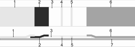
The invention discloses a bovine viral diarrhea virus differential detection test strip and a preparation method thereof, which are used for rapid and sensitive detection of BVDV antibodies. The test strip is composed of a support base plate, a coated adsorption material layer, a protein marker bearing material layer, a sample buffer material layer, a liquid absorbent material layer, and an outer protective material; the coated absorbent material is provided with a test line and a quality control line The protein label of the test strip is labeled with colloidal gold; the protein used for the protein label is the phage monoclonal combined with the BVDV antibody obtained by screening from the phage polypeptide library; the polypeptide epitope expressed by the phage monoclonal is LTPHKHHKHLHA. The preparation method comprises the preparation of gold-labeled phage monoclonal, the preparation of gold-labeled pads, the preparation of nitrocellulose membrane, and the assembly of various parts to obtain the finished product. The invention can obtain detection results within ten minutes, is fast and sensitive, has low cost, is simple to operate, and is easy to be popularized and applied in production and scientific research at the grassroots level.

Description

technical field [0001] The invention relates to the technical field of livestock virus diagnosis, in particular to a bovine viral diarrhea virus differential detection test strip and a preparation method thereof. Background technique [0002] Bovine viral diarrhea virus (BVDV), classical swine fever virus (CSFV) and human hepatitis C virus (Hepatitis C virus, HCV) all belong to the Pestivirus genus of the family Flaviviridae. [0003] BVDV infection causes immunosuppression, which stimulates respiratory tract infection, and virus infection causes abortion of pregnant cows, which causes huge economic losses to the cattle industry, and is one of the most important diseases faced by the cattle industry. BVDV can be transmitted through the placenta, resulting in persistent fetal infection (Persistently infection, PI), and PI cattle are infected throughout their lives and become an important source of infection. In addition to infecting cattle, BVDV virus can also infect more th...

Claims


Application Information

Patent Timeline
Patent Type & Authority Patents(China)
IPC IPC(8): G01N33/569G01N33/558
CPCG01N33/558G01N33/569
Inventor 刘东民朱礼倩傅小恬陈鑫烨杨继飞王丽李青梅郭军庆邢广旭周景明刘运超
Owner 河南中泽生物工程有限公司
Learn More