Tilapia brain cell line and application thereof
A tilapia and brain cell technology, applied in the field of biological cytology, can solve the problems of amplification and purification, lack of Luohu virus, inability to identify TiLV infection, etc., and achieves the effect of a simple and convenient preparation method
- Summary
- Abstract
- Description
- Claims
- Application Information
AI Technical Summary
Problems solved by technology
Method used
Image
Examples
Embodiment 1
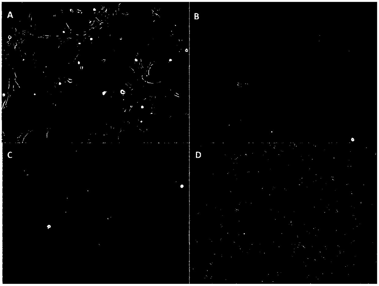
[0026] Embodiment 1 A kind of construction method of tilapia brain cell line
[0027] The above-mentioned method of the present invention is described in detail as follows according to detailed steps:
[0028] 1. Preparation of tilapia brain tissue:
[0029] Soak fresh tilapia in 75% alcohol for 1-2min, put it in an ultra-clean bench, remove the brain tissue aseptically, rinse it twice with PBS, and put it in a 5ml sterile penicillin vial (containing 5% fetal bovine serum). M199 culture medium), cut it into about 1mm with surgical scissors 3 For fragments, add 5ml PBS to collect them into a 15ml centrifuge tube, centrifuge at 1000rpm for 3 minutes, discard the supernatant after centrifuging to collect tissue fragments, and repeat once. Add collagenase type Ⅰ about 5 times the volume of the tissue block, digest at 37°C for 30 minutes, remove the digestion solution, wash twice with PBS, then add 0.25% trypsin, digest at room temperature for 6-7min, add culture medium to stop d...
Embodiment 2
[0039] The culture condition research of embodiment 2 tilapia brain cells
[0040] 1 Determination of the best culture medium for tilapia brain cells
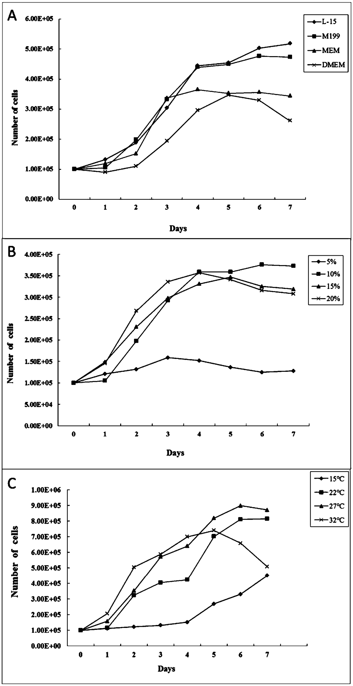
[0041] Four cell culture media, DMEM, M199, MEM, and L-15, were selected, and FBS with a final concentration of 10% was added to prepare the cell culture medium. Adjust the cell density to 4 x 10 5 mL -1 , each of the four mediums was inoculated in a 6-well plate at an amount of 2.5 mL / well, and cultured in an incubator at 27°C. Cells in 3 wells were taken out from each experimental group every 1 day, collected and counted by Trypsin-EDTA digestion method, co-cultured for 7 days, counted 7 times continuously, and the growth curve was drawn. Determine its optimum medium as M199 or L-15 medium ( figure 2 A).
[0042] 2 Determination of optimal serum concentration of tilapia brain cells
[0043] Prepare culture solutions with FBS concentrations of 5%, 10%, 15%, and 20% respectively, and adjust the cell density to 4×10 5 mL...
Embodiment 3
[0046] Example 3 Chromosome determination of tilapia brain cells
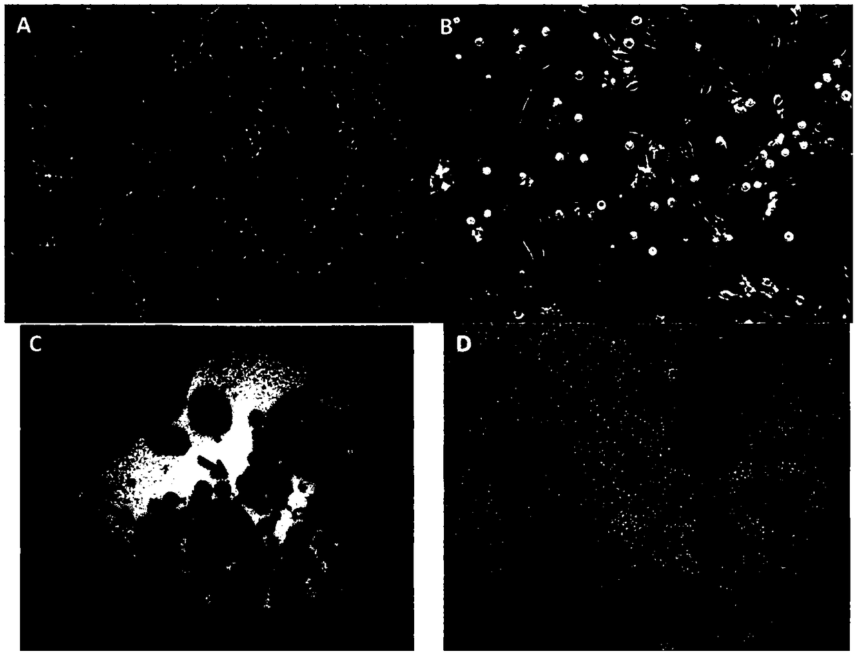
[0047] The 5th and 60th generation tilapia brain cells were added with colchicine at a final concentration of 20 μg / mL in the logarithmic growth phase, and the cells were collected after treatment at 27°C for 4 hours, treated with 0.075mol / L KCl for 25 minutes, and added 1 mL Pre-cooled Carnot's fixative, centrifuged at 1000r / min for 5min to remove the supernatant, then fixed with pre-cooled Carnot's fixative for 3 times, 15min each time. The slices were dropped by cold drop method, and after drying, stained with 5% Giemsa for 25min. For microscopic examination, 100 split phases were selected for karyotype analysis and statistics. The results show( Figure 4 ), 60% of the chromosomes of the tilapia brain cells of the 5th generation were 2n=44; 43% of the chromosomes of the tilapia brain cells of the 60th generation were 2n=50. This indicates that the cells are a permanent cell line and can proliferate indefi...
PUM
| Property | Measurement | Unit |
|---|---|---|
| Titer | aaaaa | aaaaa |
Abstract
Description
Claims
Application Information
 - R&D
- Intellectual Property
- Life Sciences
- Materials
- Tech Scout
- Unparalleled Data Quality
- Higher Quality Content
- 60% Fewer Hallucinations
Browse by: Latest US Patents, China's latest patents, Technical Efficacy Thesaurus, Application Domain, Technology Topic, Popular Technical Reports.
© 2025 PatSnap. All rights reserved.Legal|Privacy policy|Modern Slavery Act Transparency Statement|Sitemap|About US| Contact US: help@patsnap.com


