Application of anti-adhesion polypeptide, implanted medical instrument and preparation method thereof

A technology for implanting medical devices and adhesion, applied in medical science, coating, surgery, etc., can solve the problems of hyperplasia, poor specificity, etc., and achieve the effects of inhibiting inflammatory reactions, improving efficiency, and improving purity

Inactive Publication Date: 2018-12-04
SHANGHAI MICROPORT MEDICAL (GROUP) CO LTD
View PDF4 Cites 8 Cited by
  • Summary
  • Abstract
  • Description
  • Claims
  • Application Information

AI Technical Summary

Problems solved by technology

[0015] The invention provides the application of an anti-adhesion polypeptide, an implanted medical device and its preparation method, which are used to solve the problem of other cell proliferation c...

Method used


Image

  • Application of anti-adhesion polypeptide, implanted medical instrument and preparation method thereof
  • Application of anti-adhesion polypeptide, implanted medical instrument and preparation method thereof
  • Application of anti-adhesion polypeptide, implanted medical instrument and preparation method thereof

Examples


Embodiment 1

[0066] Example 1 Cell experiment

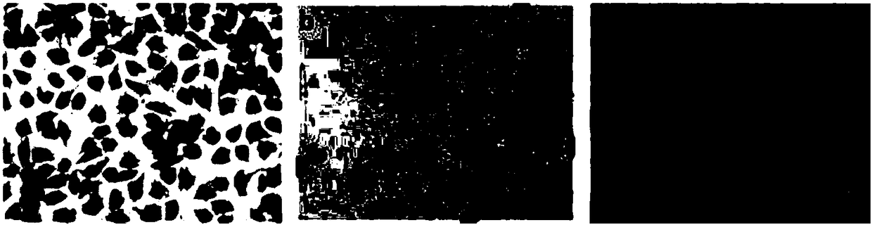
[0067] To verify the adhesion performance of the cells treated with the anti-cell adhesion polypeptide on the surface of the culture plate.

[0068] (1) Cell culture

[0069] Human arterial smooth muscle cells were cultured in DMEM (Gibico brand) high glucose medium containing 10% FBS (Gibico brand). Human umbilical vein endothelial cells cultured in EGM TM -2BulletKit TM (Lonza, CC-3162) medium containing 10% FBS and various growth factors.

[0070] (2) Cell inoculation

[0071] When the number of cells reaches 80% of the bottom area of ​​the culture flask, suck off the medium and add trypsin. When the cells shrink from the bottom of the culture flask but do not completely detach, suck off the trypsin and tap the bottom of the flask until the cells are completely detached from the bottom of the flask. Add 10 mL of PBS solution containing 1% FBS to stop the digestion, and pipette the cells to form a cell suspension. Aliquot 10mL of the...

Embodiment 2

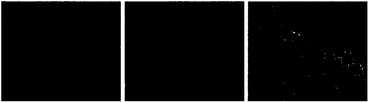
[0077] Embodiment 2 animal experiments

[0078] To verify the effect of anti-adhesion peptides on cell-trapping scaffolds to promote endothelial repair.

[0079] (1) Sample preparation

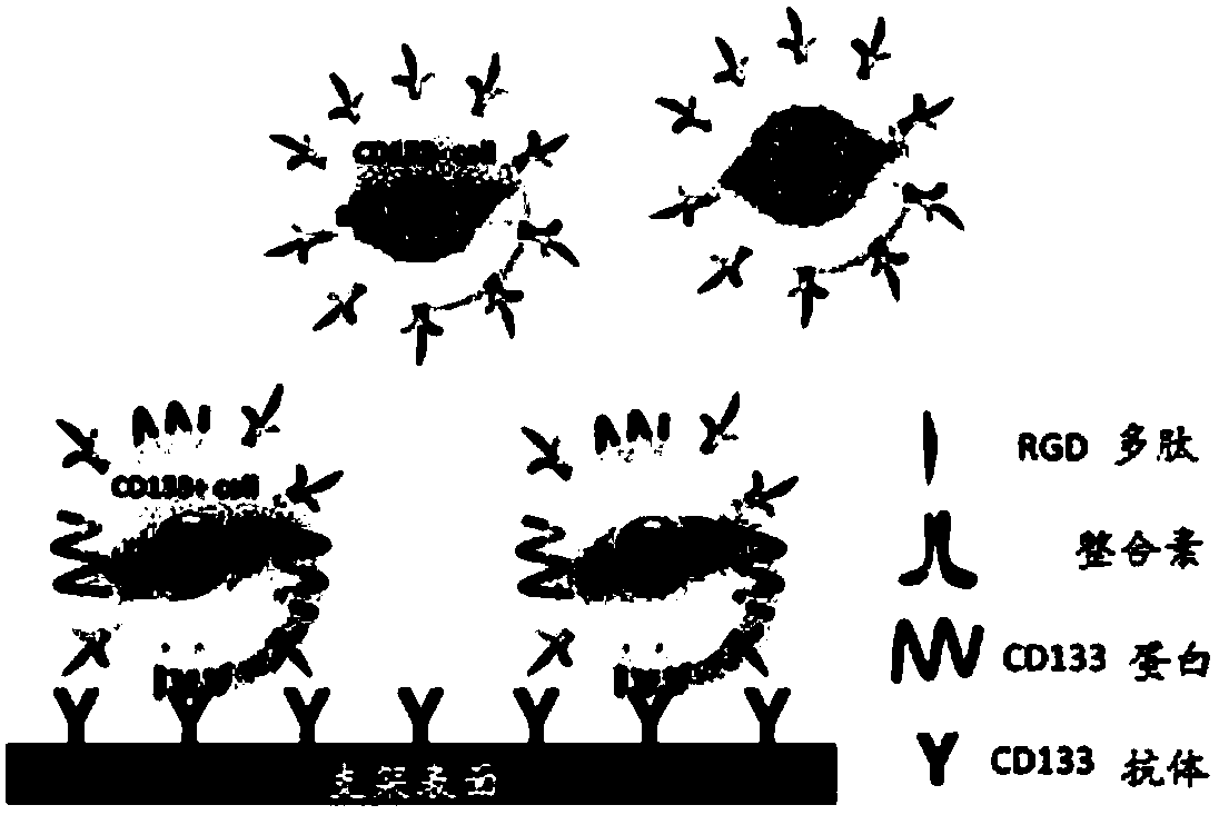
[0080] (1) Antibody loading

[0081] Prepare PEI solution (5mg / mL, 0.15M NaCl), chitosan solution (CS, 1mg / mL, 0.15M NaCl, pH=4.0), sodium hyaluronate solution (HA, 1mg / mL, 0.15M NaCl, pH =6.0), NaCl solution (0.15M), AC133 (CD133-1) antibody solution 1g / mL.

[0082] Cut the L605 bare metal bracket into a structure with a hole in the bracket rod. with H 2 SO 4 :H 2 o 2 (30%)=3:1 solution for 30 minutes, followed by deionized ultrasonic cleaning for 3 times, 10 minutes each time. After being blown dry with nitrogen gas, first dip it in polyethyleneimine (PEI) solution, let it stand for 30 minutes, take it out, wash it, and blow it dry. Soak the PEI-treated stent in sodium hyaluronate solution, wash and dry after standing for 20 minutes, and obtain the outer layer of HA; then soak the s...

Embodiment 3

[0095]Prepare PEI solution (5mg / mL, 0.15M NaCl), chitosan solution (CS, 1mg / mL, 0.15M NaCl, pH=4.0), sodium hyaluronate solution (HA, 1mg / mL, 0.15M NaCl, pH =6.0), NaCl solution (0.15M), cell capture substance AC133 (CD133-1) antibody solution 10 mg / mL.

[0096] Cut the L605 bare metal bracket into a structure with a hole in the bracket rod. with volume ratio H 2 SO 4 :H 2 o 2 (30%) = 3:1 solution washing for 30 minutes, washing with pure water, and drying with nitrogen. Then soak it in PEI solution, take it out after 30 minutes, wash and dry it. Soak the PEI-treated stent in sodium hyaluronate solution, wash and dry after standing for 20 minutes, and obtain the outer layer of HA; then soak the stent in chitosan solution, wash and dry after standing for 20 minutes, The CS outer layer was obtained; the above process was repeated 7 times to obtain a sodium hyaluronate / chitosan self-assembled 7-layer film. Soak the scaffold in the CD133-1 (AC133) antibody solution, and let...


PUM


Abstract

The invention belongs to the technical field of medical apparatus and instruments, and specially relates to the technical field capable of performing damage endothelium repair on a device implantationposition. The invention provides an application of anti-adhesion polypeptide in preparation of an implanted medical instrument. The implanted medical device is loaded with a cell trapping substance and the anti-adhesion polypeptide. A preparation method of the implanted medical instrument includes the following steps: loading the cell trapping substance on a surface of the cell trapping substancethat the implanted medical device plan to load, and loading the anti-adhesion polypeptide into a hole or a groove of the implanted medical device. According to the technical scheme, from the perspective of anti-cell adhesion, a mechanism of combining the anti-adhesion peptide with an adhesion molecule on a cell membrane is adopted, and the adhesion of other types of cells except captured specialcells to the surface of the implanted medical device is blocked, and therefore, the purity of the captured cells can be greatly improved, the efficiency of endothelial repair is improved, thereby achieving the purpose of promoting endothelial neogenesis, inhibiting intimal hyperplasia, and inhibiting an inflammatory reaction.

Description

technical field [0001] The invention belongs to the technical field of medical devices, and in particular relates to a technical field capable of repairing damaged endothelium at device implantation sites. Background technique [0002] During the implantation of medical devices such as stents and occluders into the human body, the endothelial tissue around the implanted tissue will be damaged to a certain extent due to axial shear force and friction force. The damage of this endothelial tissue will cause the surrounding tissues to suffer from inflammatory reactions, intimal hyperplasia and other hazards, and long-term endothelial damage will cause serious sequelae such as vascular restenosis and thrombosis. Therefore, it is necessary to repair the damaged endothelial tissue. [0003] In recent years, people have tried a variety of methods for endothelial repair. The main technical point is to capture endothelial cells, endothelial progenitor cells, endothelial precursor cel...

Claims


Application Information

Patent Timeline
IPC IPC(8): A61L31/04A61L31/16A61L31/06A61L31/08A61L31/14
CPCA61L31/047A61L31/06A61L31/08A61L31/088A61L31/14A61L31/16A61L2300/252A61L2300/258A61L2300/41A61L2300/412C08L71/02
Inventor 王玄陈陆李俊菲
Owner SHANGHAI MICROPORT MEDICAL (GROUP) CO LTD
Why Eureka
Try Eureka