Oligo probe and preparation method and application thereof

A probe and reaction technology, applied in the field of Oligo probe and its preparation, can solve the problems of increasing complexity, dependence, and hindering probe preparation, and achieve the effect of low hybridization background, high specificity and high specificity

Active Publication Date: 2018-12-14
中山康源基因技术科技有限公司
View PDF4 Cites 1 Cited by
  • Summary
  • Abstract
  • Description
  • Claims
  • Application Information

AI Technical Summary

Problems solved by technology

This method relies heavily on the probe sequence, and it is hindered to extend to the preparation of probes for a large number of genes.
[0006] Secondly, relative to the synthetic method of making probes, the probe sequence can be specifically selected. Traditional nucleic acid probes also have the problem of repetitive s

Method used


Image

  • Oligo probe and preparation method and application thereof
  • Oligo probe and preparation method and application thereof
  • Oligo probe and preparation method and application thereof

Examples


Embodiment 1

[0059] HER2, also known as HER2 / neu, c-erbB-2, is a member of the epidermal growth factor receptor (EGFR) family. Studies have shown that HER2 gene amplification exists in more than 30% of human tumors, such as breast cancer, ovarian cancer, Gastric cancer and prostate cancer etc. Breast cancer is a common tumor that threatens women's life and health, and 20%-30% of primary invasive breast cancers have HER2 gene amplification. Fluorescence in situ hybridization (FISH) is a common way to detect HER2 gene amplification.

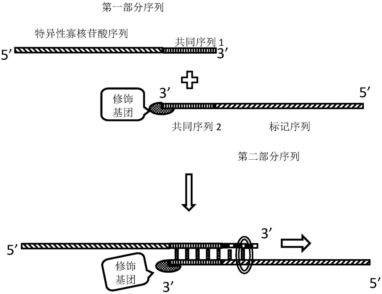
[0060] (1) Design of the probe

[0061] Find the HER2 gene sequence on chromosome 17 from NCBI, remove the repetitive gene sequence, truncate it into a probe sequence with a length of 50-150bp, and add a common sequence to the 3' end of each sequence (5'—TGGTCGG— 3').

[0062] Probe 1: 5'—ggatccctga tggggagaat gtgaaaattc cagtggccat caaagtgttgTGGTCGG—3';

[0063] Probe 2: 5'—agggaaaaca catcccccaa agccaacaaa gaaatcttag acgtaagcccctccaccctc tcctgctagg TGGTCGG—...

Embodiment 2

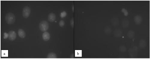
[0095] The above-mentioned labeled HER2 probe (Cy3-labeled HER2 probe) is used for hybridization of cell smears, figure 1 It is a schematic diagram of the labeled probe, which enters the smear cells under the action of the hybridization buffer and specifically binds to the chromosome. The fluorescein combined with the labeled deoxynucleotide emits fluorescence of a specific wavelength under the excitation light. Specific fluorescent signals can be seen after the corresponding grating, such as image 3 , which is the specific bright spot of the excited probe in the cell. This embodiment comprises the steps:

[0096] (1) Preparation of cell smears

[0097] Add colchicine to the culture dish of human embryonic kidney 293T cells cultured in vitro (Shanghai Cell Bank, Chinese Academy of Sciences) to a final concentration of 0.4 μg / mL, and treat at 37°C for 1.5h. Add 25%wt trypsin solution, digest at 37°C for 2min, add 10mL of potassium chloride solution with a concentration of 0...

Embodiment 3

[0107] The 293T cell line in Example 2 was replaced by the HER-2 positive Skbr-3 cell line (Shanghai Cell Bank, Chinese Academy of Sciences), and the cell droplet and hybridization and cleaning methods were the same as in Example 2, and the hybridization results were as follows: Figure 4 , the results showed that the HER2 gene copy number increased significantly in the HER-2 positive Skbr-3 cell line, and the probe prepared by the invention can effectively distinguish the HER-2 positive tumor cell line.


PUM


Abstract

The invention discloses an Oligo probe and a preparation method and application thereof. The preparation method comprises: mixing a first partial sequence and a second partial sequence, carrying out denaturation and annealing in a labeled deoxyribonucleotide system, and adding a DNA polymerase into the system for a polymerization reaction to obtain the Oligo probe. The first partial sequence is acommon sequence 1 connected to the end 3' of a specific oligonucleotide sequence and the second partial sequence is a common sequence 2 connected to the end 3' of a labeled sequence. The specific oligonucleotide sequence is specifically bonded to a gene to be detected and does not contain the repeated sequence. The common sequence 2 is a sequence with a modified end 3' and the common sequence 1 iscomplementary with the common sequence 2. The Oligo probe has high sensitivity and specificity and can be used for preparation of a FISH probe. The FISH probe easily enters cells and has a fast hybridization reaction rate. Through a specific hybridization buffer, the hybridization time can be shortened to 1 to 2h.

Description

technical field [0001] The invention relates to a nucleic acid probe preparation and labeling method for FISH technology, in particular to an Oligo probe and its preparation method and application. Background technique [0002] By using nucleic acid probes, such as DNA or RNA probes with a certain label, to detect and display abnormalities in chromosome structure and gene expression at the cellular or chromosome level, this is a very intuitive way to show many problems at the genetic level of living organisms. This visualization technique is called in situ hybridization (ISH), and the probe labeling method used can be roughly divided into "radioisotope labeling method" and "fluorescein labeling method". Fluorescence in situ hybridization (FISH), which has been gradually developed for the application of needles, has the characteristics of economy, safety, speed, and stability. , tumor genetics and genome evolution research and many other fields. The principle is to use a nu...

Claims


Application Information

Patent Timeline
IPC IPC(8): C12Q1/6806C12Q1/6841
CPCC12Q1/6806C12Q1/6841C12Q2521/101C12Q2563/107
Inventor 蔡伟文邓佳颜飞地
Owner 中山康源基因技术科技有限公司
Why Eureka
Try Eureka