Kit for detecting HPV (Human Papillomavirus)
A kit and sequence technology, applied in the field of bioengineering, can solve the problems of low specificity and sensitivity, and achieve the effects of improving sensitivity and specificity, shortening reaction time, and improving detection efficiency
- Summary
- Abstract
- Description
- Claims
- Application Information
AI Technical Summary
Problems solved by technology
Method used
Image
Examples
Embodiment 1
[0045] Embodiment 1 is used to detect the test kit of HPV
[0046] A kit for detecting HPV based on a recombinant high-sensitivity amplification method, comprising:
[0047] A first primer pair (SEQ ID NO.1 and SEQ ID NO.2) for amplifying HPV16 and a second primer pair (SEQ ID NO.3 and SEQ ID NO.4) for amplifying HPV18; Uvs X (SEQ ID NO.5), Uvs Y (SEQ ID NO.6), SSB (SEQ ID NO.7) and DNA polymerase (SEQ ID NO.8); and amplification buffer: 20mM Tris-HCl, 10mM (NH 4 ) 2 SO 4 , 50mM KCl, 2mM MgSO 4 and 0.1% 20 (pH=8.8).
Embodiment 2D
[0048] Example 2 DNA extraction (extraction method)
[0049] The extraction of DNA in the detection sample includes the following steps:
[0050] 1. Add 1ml of extraction buffer (100mM Tris-HCl, pH=8.0; 50mM EDTA, pH=8.0; 500mM NaCl, +0.07% mercaptoethanol) to cervical tissue (Cervical tissue) cells, and mix thoroughly.
[0051] 2. Add 130ul 10% SDS and a small amount of proteolytic enzyme K, shake it upside down several times, and place it in a water bath at 65°C for 1 hour.
[0052] 3. Add 300ul 5M potassium acetate (potassium acetate), mix thoroughly, and then place on ice for 30 minutes to facilitate protein precipitation.
[0053] 4. Centrifuge at 12000 rpm for 30 minutes, take out the supernatant and transfer to a new centrifuge tube (eppendorf tube).
[0054] 5. Add twice the volume of isopropanol and centrifuge at 12000rpm for 30 minutes.
[0055] 6. Pour off the supernatant, add 70% ethanol, and centrifuge at 12000 rpm for 30 minutes.
[0056] 7. Pour off the supern...
Embodiment 3
[0059] Embodiment 3HPV detects
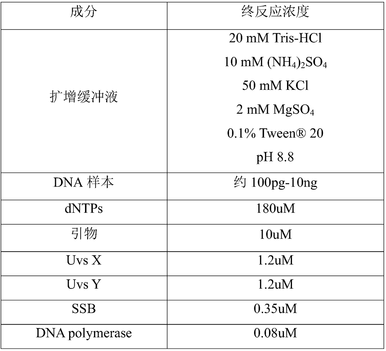
[0060] The kit in Example 1 and the extracted DNA sample in Example 2 were added to the micro test tube (final volume: 50ul) according to the proportions in Table 1 below, and shaken homogeneously in an environment of 37-40°C React at 300rpm for 10-20 minutes.
[0061] Table 1
[0062]
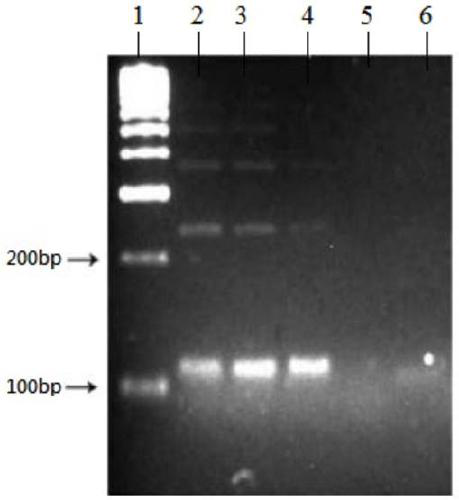
[0063] After the reaction is finished, the reaction product is electrophoresed in 3-4% agaric gum, and the size of the DNA product is judged (adding a tracking dye, performing contrast electrophoresis with the control group, and putting it into a DNA marker for comparison). The power plug (note the position of the positive and negative poles) is connected to the power supply. During the electrophoresis process, the negatively charged DNA molecules will move from the negative pole to the positive pole, and run the gel in 100vol for about 10 minutes. Put the agaric gum on the strong UV transmission light source plate with gloves on, and then take a photo of ...
PUM
 Abstract
Description
Claims
Application Information
 - R&D
- Intellectual Property
- Life Sciences
- Materials
- Tech Scout
- Unparalleled Data Quality
- Higher Quality Content
- 60% Fewer Hallucinations
Browse by: Latest US Patents, China's latest patents, Technical Efficacy Thesaurus, Application Domain, Technology Topic, Popular Technical Reports.
© 2025 PatSnap. All rights reserved.Legal|Privacy policy|Modern Slavery Act Transparency Statement|Sitemap|About US| Contact US: help@patsnap.com


