
One-step PCR detection of avian leukosis virus a/b/j/k subgroup primer set and kit
A technology of avian leukosis virus and primer set, applied in the field of molecular biology, can solve the economic loss of poultry industry and the lack of ALV vaccine and other problems, achieve simple and rapid typing detection, reduce detection time and cost, and have good specificity
- Summary
- Abstract
- Description
- Claims
- Application Information
AI Technical Summary
Problems solved by technology
Method used
Image

Examples
Embodiment 1
[0038] The design of embodiment 1 PCR primer
[0039]Download the full-length ALV-A, ALV-B, ALV-J, and ALV-K gene sequences in Table 1 from the GenBank database, use DNAstar software for sequence alignment, and design a line in the most conserved pol gene region common to the four subgroups The shared upstream primer P1, four different downstream primers P2, P3, P4 and P5 were designed in the subgroup-specific env gene region. Among them, P1 and P2 can amplify a 375 bp fragment for detecting ALV-A, P1 and P3 can amplify a 581 bp fragment for detecting ALV-B, and P1 and P4 can amplify a 683 bp fragment For detecting ALV-J, P1 and P5 can amplify a 1377 bp fragment for detecting ALV-K. Primer sequences are listed below in Table 1.
[0040] Table 1
[0041] Primer Subtype 5'-3' sequence product size P1 GCCAAACGGATTTCTGCCTT P2 ALV-A AACCCAGATACCAAGGAACC 375 bp P3 ALV-B ACAGATGGACCAATTCTGTCTC 581 bp P4 ALV-J ATTGTTCCACAAACACCT...
Embodiment 2
[0042] Example 2 Standard positive plasmid construction
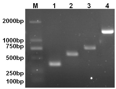
[0043] The specific bands amplified from ALV-A, ALV-B, ALV-J and ALV-K proviral DNA were recovered with a DNA gel extraction kit, cloned into pMD-18-T vectors, and transformed into DH5α Escherichia coli is competent, and the single clone is picked and the plasmid is extracted and sent for sequencing identification. The result is as figure 1 As shown, M: DNA molecular standard DL2000; 1: pMD-A; 2: pMD-B; 3: pMD-J; 4: pMD-K, where the plasmid for cloning the 375 bp fragment of ALV-A is named pMD-A , is the standard positive control plasmid for ALV-A subtype virus; the plasmid for cloning the 581 bp fragment of ALV-B is named pMD-B, the standard positive control plasmid for ALV-B subtype virus; the plasmid for cloning the 683 bp fragment of ALV-J is named pMD-J, ALV-J subtype virus standard positive control plasmid; the plasmid for cloning the ALV-K1377 bp fragment was named pMD-K, ALV-K subtype virus standard positive c...
Embodiment 3
[0044] Example 3 PCR method establishment and condition optimization
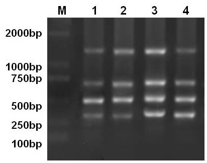
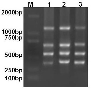
[0045] The extracted ALV-A, ALV-B, ALV-J and ALV-K proviral DNA were diluted to 100 ng / μl respectively, and mixed at equal concentrations to serve as templates. In a 50 μl reaction system, the primer concentration was increased from 10 pmol / L ( The upstream primer P1 is 2 μl, 4 μl, 6 μl and 8 μl respectively; the downstream primers P2, P3, P4 and P5 are each 2 μl (the results are as follows figure 2 Shown M: DNA molecular standard DL2000; 1: 2 μL upstream primer; 2: 4 μL upstream primer; 3: 6 μL upstream primer; 4: 8 μL upstream primer)), annealing temperature (53 ℃, 56 ℃, 59 ℃ ( The result is as image 3 The shown M: DL 2000 standard molecular weight; 1-3: the amplification results when the annealing temperature is 53°C, 56°C, and 59°C respectively)) and other conditions were optimized, and the optimal conditions for screening and obtaining multiplex PCR reactions are shown in Table 2 below :
[0046] ...
PUM
 Abstract
Description
Claims
Application Information