Fluorescent biosensor for detecting triphosadenine and preparation method thereof
A biosensor, adenosine triphosphate technology, applied in the direction of fluorescence/phosphorescence, instruments, measuring devices, etc., can solve the problems of long detection cycle, low specificity and sensitivity, etc., and achieve the effect of short detection cycle, easy operation and reduced complexity
- Summary
- Abstract
- Description
- Claims
- Application Information
AI Technical Summary
Problems solved by technology
Method used
Image
Examples
Embodiment 1
[0056] Example 1 Preparation of Circular Template and Composite Probe
[0057] Prepared with 50 mM Tris-HCl, 10 mM MgCl 2 , T4 DNA Ligase Reaction Buffer with 10 mM DTT and 1 mM ATP. Formulated with 10 mM Na 2 HPO 4 , 10 mM NaH 2 PO 4 , 140 mM NaCl, 1 mM KCl, 1 mM MgCl 2 , 1mM CaCl 2 , PBS buffer solution of pH=7.4.
[0058] (1) Mix 42 μL sterile water, 6 μL linear template (10 μM), 6 μL ligation probe (10 μM) and 6 μL 10× T4 DNA ligase buffer, denature at 95°C for 5 min, then Slowly cool down to room temperature to complete the hybridization, then add 3 μL of T4 DNA ligase (60 U / μL) to the reaction system, and react at 16°C for 20 hours; after that, the reaction system is placed in a water bath at 65°C for 15 minutes , inactivate the T4 DNA ligase in the system.
[0059] (2) Add 3 μL of exonuclease Ⅰ (20 U / μL) and 3 μL of exonuclease Ⅲ (100 U / μL) to the above reaction system and react at 37°C for 2 h; then put the reaction system at 85°C Heated in a water bath for 1...
Embodiment 2
[0062] A method for preparing a fluorescent biosensor of the present invention, comprising the following steps:
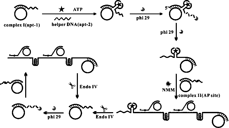
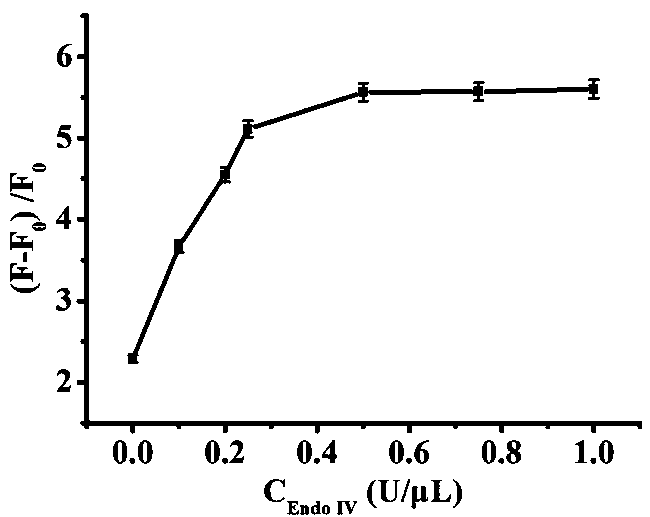
[0063] (1) Mix 2 μL composite probe I (1 μM), 2 μL dNTP (1 mM), 2 μL composite probe II (1 μM), 2 μL Apt-2 (1 μM), 2 μL phi29 DNA polymerase (1 U / μL), 2 μL endonuclease IV (concentrations are 0.1 U / μL, 0.2U / μL, 0.25 U / μL, 0.5 U / μL, 0.75 U / μL, 1 U / μL), 2 μL NMM (2.4 μM) in 2 μL buffer (50 mMTris-HCl, 10 mM MgCl 2 , 10 mM (NH 4 ) 2 SO 4 , 4 mM DTT, pH 7.5) after mixing, add ATP (100 nM) respectively, mix and react at a constant temperature of 37°C for 90 min;
[0064] (3) Dilute the solution obtained in step (2) with water to 100 μL, and then perform fluorescence detection; the excitation wavelength is set to 486 nm, the emission wavelength is 610 nm, and the detection range is 560 nm-640 nm, and the fluorescence signal changes are read.
[0065] See the test results figure 2 , it can be seen from the figure that as the amount of endonuclease IV increases, the...
Embodiment 3
[0069] A method for preparing a fluorescent biosensor of the present invention, comprising the following steps:
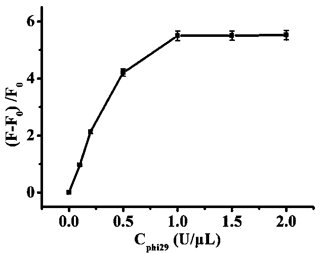
[0070] (1) Mix 2 μL composite probe I (1 μM), 2 μL dNTP (1 mM), 2 μL composite probe II (1 μM), 2 μL Apt-2 (1 μM), 2 μL phi29 DNA polymerase (0.1 U / μL, 0.2 U / μL, 0.25 U / μL, 0.5 U / μL, 1 U / μL, 1.5 U / μL, 2 U / μL), 2 μL endonuclease IV (0.5 U / μL μL), 2 μL NMM (2.4 μM) in 2 μL buffer (50 mM Tris-HCl, 10 mM MgCl 2 , 10 mM (NH 4 ) 2 SO 4 , 4 mM DTT, pH 7.5) after mixing, add ATP (100 nM) respectively, mix and react at a constant temperature of 37°C for 90 min;
[0071] (3) Dilute the solution obtained in step (2) with water to 100 μL, and then perform fluorescence detection; the excitation wavelength is set to 486 nm, the emission wavelength is 610 nm, and the detection range is 560 nm-640 nm, and the fluorescence signal changes are read.
[0072] See the test results image 3 , it can be seen from the figure that as the amount of phi29 DNA polymerase increases, the ...
PUM
 Abstract
Description
Claims
Application Information
 - R&D
- Intellectual Property
- Life Sciences
- Materials
- Tech Scout
- Unparalleled Data Quality
- Higher Quality Content
- 60% Fewer Hallucinations
Browse by: Latest US Patents, China's latest patents, Technical Efficacy Thesaurus, Application Domain, Technology Topic, Popular Technical Reports.
© 2025 PatSnap. All rights reserved.Legal|Privacy policy|Modern Slavery Act Transparency Statement|Sitemap|About US| Contact US: help@patsnap.com


