Method of establishing chronic ocular hypertension animal model

A technology of animal model and establishment method, which is applied in medical science, veterinary instruments, veterinary surgery, etc., can solve problems such as short maintenance time, unstable intraocular pressure increase, and increased intraocular pressure, achieving increased time and reduced Effect of aqueous humor outflow and reflux prevention

Active Publication Date: 2019-03-19
SHENYANG EYE IND TECH INST LTD +1
View PDF2 Cites 6 Cited by
  • Summary
  • Abstract
  • Description
  • Claims
  • Application Information

AI Technical Summary

Problems solved by technology

[0003] At present, the more commonly used animal models of ocular hypertension include: (1) posterior chamber injection of α-chymotrypsin or carbomer ocular hypertension model: the disadvantage of this model is that it can cause more obvious inflammatory reactions, and the intraocular pressure rises suddenly , with a short maintenance time and low success rate, it is easy to cause many complications (such as lens opacity, dislocation, anterior chamber exudation, corneal edema and lens atrophy, etc.); (2) Anterior chamber injection of sodium hyaluronate high intraocular pressure model: The disadvantage of this model is that repeated injections are required to maintain high intraocular pressure, and the increase in intraocular pressure is not obvious. Usually, simple injection of sodium hyaluronate into the anterior chamber can only maintain high intraocular pressure for 5 days. Repeated injections of sodium hyaluronate are required every 5 days; (3) Laser photocoagulation of the sclera or laser photocoagulation of trabecular meshwork high intraocular pressure model: the disadvantages of this model are difficult operation, high cost, and maintenance of high intraocular pressure If the time is short, repeated laser photocoagulation is required to maintain high intraocular pressure for a long time, but it is easy to cause intraocular congestion and inflammatory reactions; (4) Anterior chamber injection of polystyrene microspheres to establish a chronic high intraocular pressure model: the The disadvantage of the model is that the distribution of polystyrene microspheres in the angle of the chamber is uneven, resulting in an unstable increase in intraocular pressure, and repeated injections of microspheres are required to maintain high intraocular pressure for a long time

Method used


Image

  • Method of establishing chronic ocular hypertension animal model
  • Method of establishing chronic ocular hypertension animal model
  • Method of establishing chronic ocular hypertension animal model

Examples


Embodiment 1

[0030] Taking rats fed for 8 weeks as an example, a chronic high intraocular pressure model was established, the right eye was used as the animal model eye, and the left eye was used as the normal control eye without any treatment.

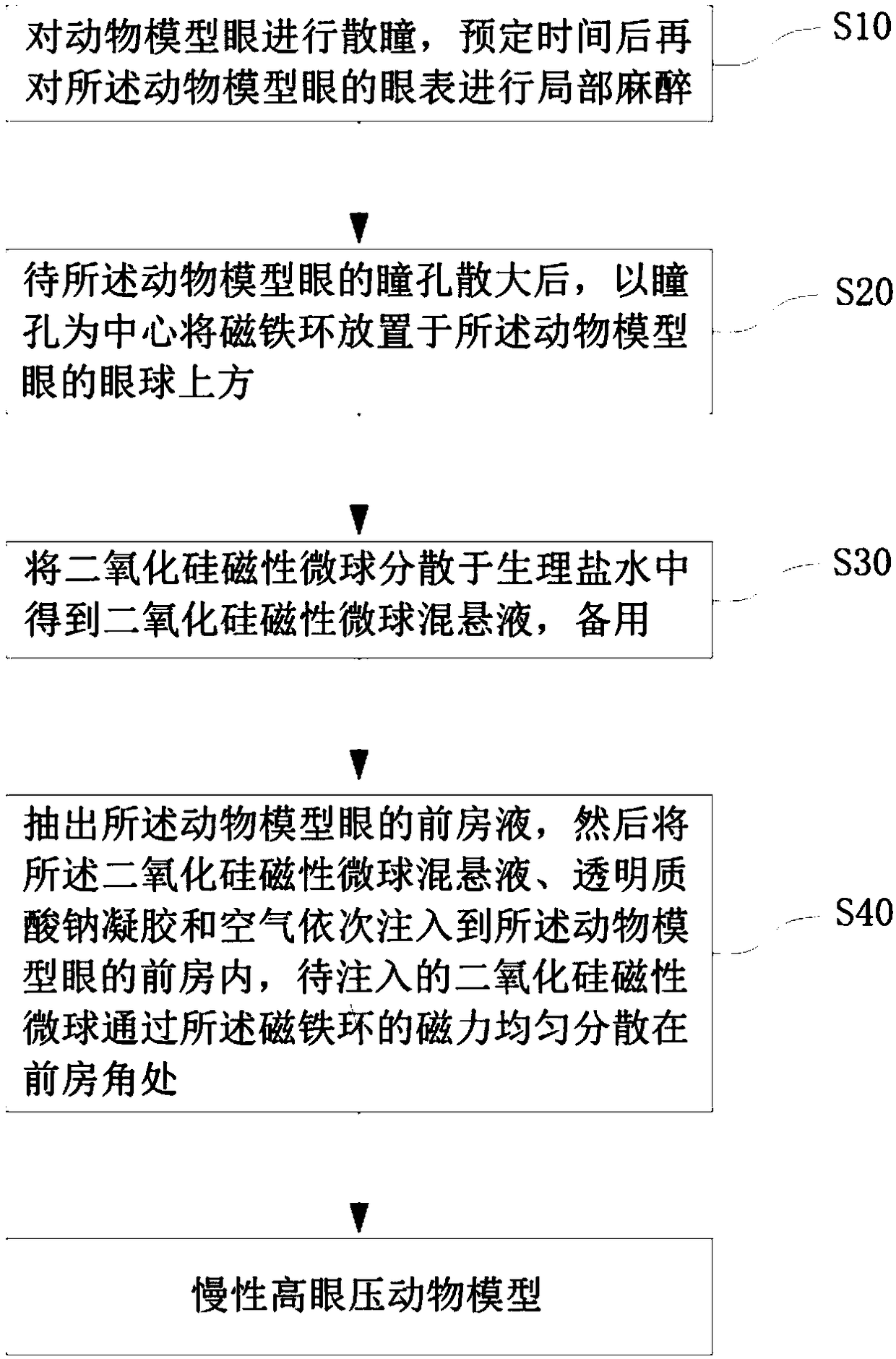
[0031] refer to figure 1 , this embodiment 1 provides a method for establishing an animal model of chronic ocular hypertension, comprising:

[0032] In step S10, dilate the animal model eye, and then perform local anesthesia on the ocular surface of the animal model eye after a predetermined time.

[0033] As an example, add compound tropicamide eye drops to the rat animal model eye (right eye) after general anesthesia for mydriasis, and then add oxybucaine hydrochloride eye drops for local anesthesia on the ocular surface after 10 minutes .

[0034] It should be understood that various feasible anesthesia methods can be used to perform general anesthesia on the rat animal model, which is not limited in the present invention.

[0035] In step S...

Embodiment 2

[0060] Taking rats fed for 8 weeks as an example, a chronic high intraocular pressure model was established, with the right eye as the model eye, and the left eye without any treatment as the normal control eye.

[0061] This embodiment 2 provides a method for establishing an animal model of chronic ocular hypertension, including:

[0062] (1) Add compound tropicamide eye drops to the rat model eye (right eye) after general anesthesia for mydriasis, and then add oxybucaine hydrochloride eye drops for local anesthesia 10 minutes later.

[0063] (2) After the pupil of the model eye is dilated, the prepared magnet ring: outer diameter 13mm, inner diameter 10mm, thickness 2mm, central magnetic field 3000Gs, is set on the orbit of the animal model eye with the pupil as the center, so that The magnet ring is placed above the eyeball.

[0064] (3) Disperse silica magnetic microspheres with a diameter of 10 μm and a magnetic strength of 50 emu / g in physiological saline to prepare a s...

Embodiment 3

[0067] Taking rats fed for 8 weeks as an example, a chronic high intraocular pressure model was established, with the right eye as the model eye, and the left eye without any treatment as the normal control eye.

[0068] This embodiment 3 provides a method for establishing an animal model of chronic ocular hypertension, including:

[0069] (1) Add compound tropicamide eye drops to the rat model eye (right eye) after general anesthesia for mydriasis, and then add oxybucaine hydrochloride eye drops for local anesthesia 10 minutes later.

[0070] (2) After the pupil of the model eye is dilated, put the prepared magnet ring: outer diameter 15mm, inner diameter 10mm, thickness 5mm, center magnetic field 3500Gs, on the orbit of the model eye with the pupil as the center, so that all The magnet ring is placed above the eyeball.

[0071] (3) Disperse silica magnetic microspheres with a diameter of 10 μm and a magnetic strength of 70 emu / g in physiological saline to prepare a suspensi...


PUM


Abstract

The invention relates to a method of establishing a chronic ocular hypertension animal model. The method includes: dilating pupils of eyes of an animal model, and after a predetermined time, subjecting the ocular surface of the animal model to local anesthesia; after the pupils of the eyes of the animal model are dilated, placing magnet rings above the eyeballs of the animal model by centering onthe pupils; dispersing silica magnetic microspheres in normal saline to obtain silica magnetic microsphere suspension for use; extracting anterior chamber fluid from the eyes of the animal model, andinjecting the silica magnetic microsphere suspension, sodium hyaluronate gel and air sequentially into the anterior chambers of the eyes of the animal model, and after the silica magnetic microspheresinjected are dispersed magnetically and evenly to the anterior chamber corners through the magnet rings, acquiring the chronic ocular hypertension animal model. The method provided herein helps evidently extend the ocular hypertension maintenance time and attain the stable and effective chromonic ocular hypertension animal model.

Description

technical field [0001] The invention belongs to the technical field of establishing animal experimental models, and in particular relates to a method for establishing an animal model of chronic high intraocular pressure. Background technique [0002] Glaucoma is a group of diseases characterized by optic nerve pitting atrophy and visual field defect, and its main risk factor is the increase of pathological intraocular pressure IOP (Intraocular Pressure). The main cause of glaucoma blindness is: the oppression and damage of high intraocular pressure on intraocular tissues, especially the ONH (Optic Nerve Head) in the posterior segment of the eye, resulting in optic atrophy and visual field loss. At present, the most important treatment for glaucoma is to reduce intraocular pressure through drugs or surgery to prevent or delay the progression of the disease. Therefore, in the field of glaucoma research, animal models of ocular hypertension play an important role in better und...

Claims


Application Information

Patent Timeline
Patent Type & Authority Applications(China)
IPC IPC(8): A61D1/00A61D7/00
CPCA61D1/00A61D7/00
Inventor 张东蕾安元龙何向东何伟
Owner SHENYANG EYE IND TECH INST LTD
Why Eureka
Try Eureka