Use of fat emulsion as photoacoustic imaging contrast agent

A fat emulsion and photoacoustic imaging technology, which is applied in the fields of biology and optics, can solve the problems of unknown biological safety, complex preparation method, weak contrast, etc., and achieves the enhancement of tissue photoacoustic imaging contrast, the preparation method is simple, and the cost is low. Effect

Inactive Publication Date: 2019-03-29
SOUTHERN MEDICAL UNIVERSITY
View PDF3 Cites 0 Cited by
  • Summary
  • Abstract
  • Description
  • Claims
  • Application Information

AI Technical Summary

Problems solved by technology

However, some inherent chromophores in the body, such as hemoglobin, have weak absorption in the near-infrared band, resulting in weak contrast inside the tissue. Therefore, research on photoacoustic imaging contrast agents has attracted a lot of attention in recent years.
The existing photoacoustic imaging contrast agents including indocyanine green and nano-gold particles have obvious photoacoustic imaging enhancement effect, but their biological safety is still unknown, and the preparation method is complicated and the cost is high.

Method used


Image

  • Use of fat emulsion as photoacoustic imaging contrast agent
  • Use of fat emulsion as photoacoustic imaging contrast agent
  • Use of fat emulsion as photoacoustic imaging contrast agent

Examples


Embodiment 1

[0025] The fat emulsion is used as a photoacoustic imaging contrast agent, and the fat emulsion enhances the photoacoustic signal of the tissue and the contrast between the tissue and the background. The fat emulsion consists of soybean oil, lecithin and glycerin, and every 1000ml of 20% fat emulsion contains 200g soybean oil, 12g lecithin and 22g glycerin. Fat emulsions are often used as supplementary drugs for intravenous nutrition in clinical practice, and have a high degree of biological safety.

[0026] Fat emulsions are used for animal or human imaging. When the fat emulsion was used for animal imaging, 20ml / kg of the fat emulsion was injected into the animal through the tail vein. When fat emulsions are used for human imaging, the fat emulsions are injected intravenously into the body.

[0027] The wavelength range of the laser used in photoacoustic imaging is in the near-infrared band. Preferably, the wavelength range of the laser used for photoacoustic imaging is i...

Embodiment 2

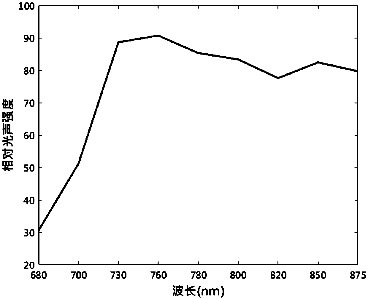
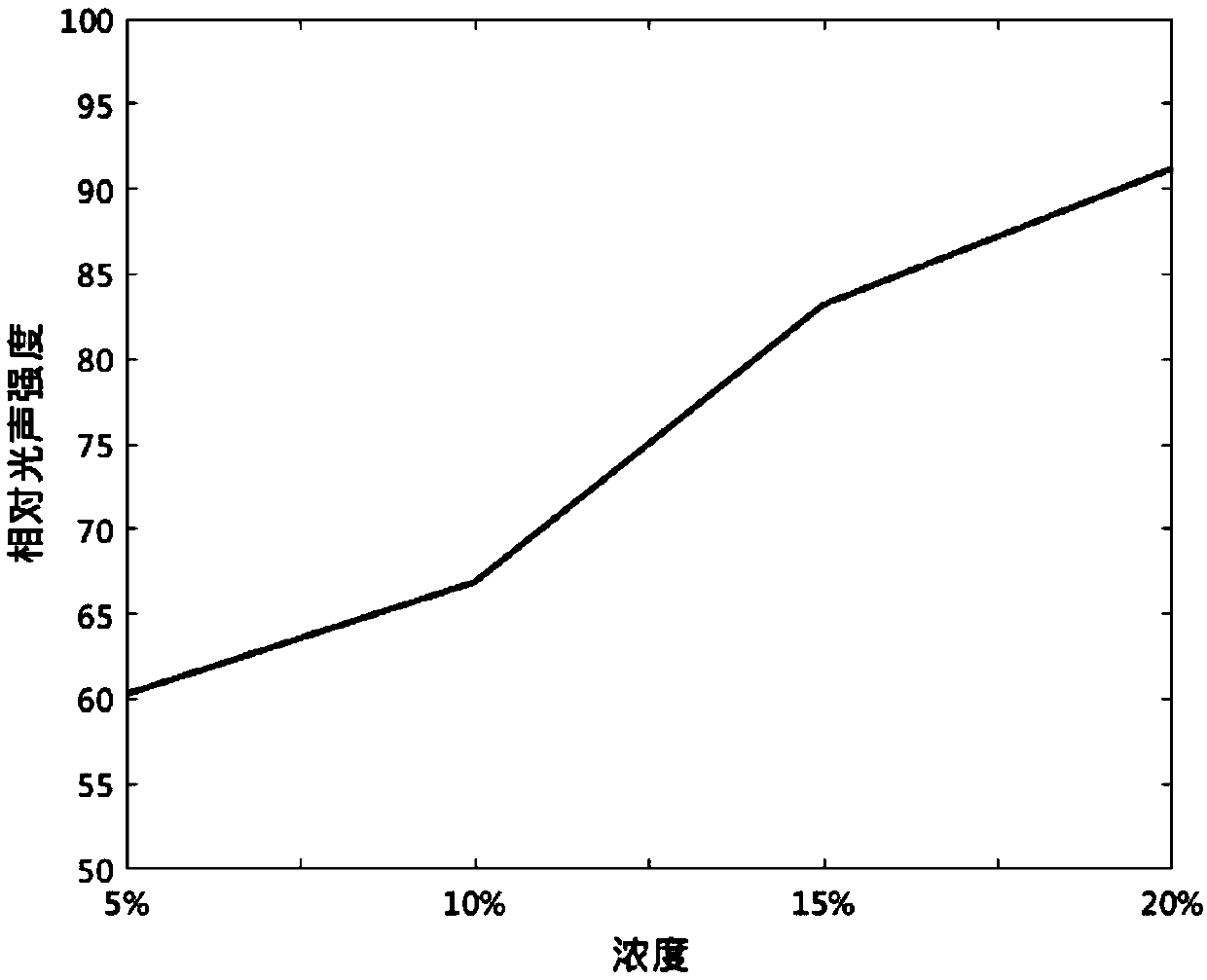
[0031] Dilute the 20% medical fat emulsion injection to 15%, 10%, 5% with normal saline, and check their photoacoustic effects under 680nm, 700nm, 730nm, 760nm, 780nm, 800nm, 820nm, 850nm, 875nm pulsed laser Signal. The results show that the fat emulsion has the strongest photoacoustic signal under 760nm laser irradiation, such as figure 1 As shown, and the photoacoustic signal intensity shows a linear increase with the increase of the fat emulsion concentration, as figure 2 As shown, this proves that the fat emulsion has the potential as a contrast agent for photoacoustic imaging.

Embodiment 3

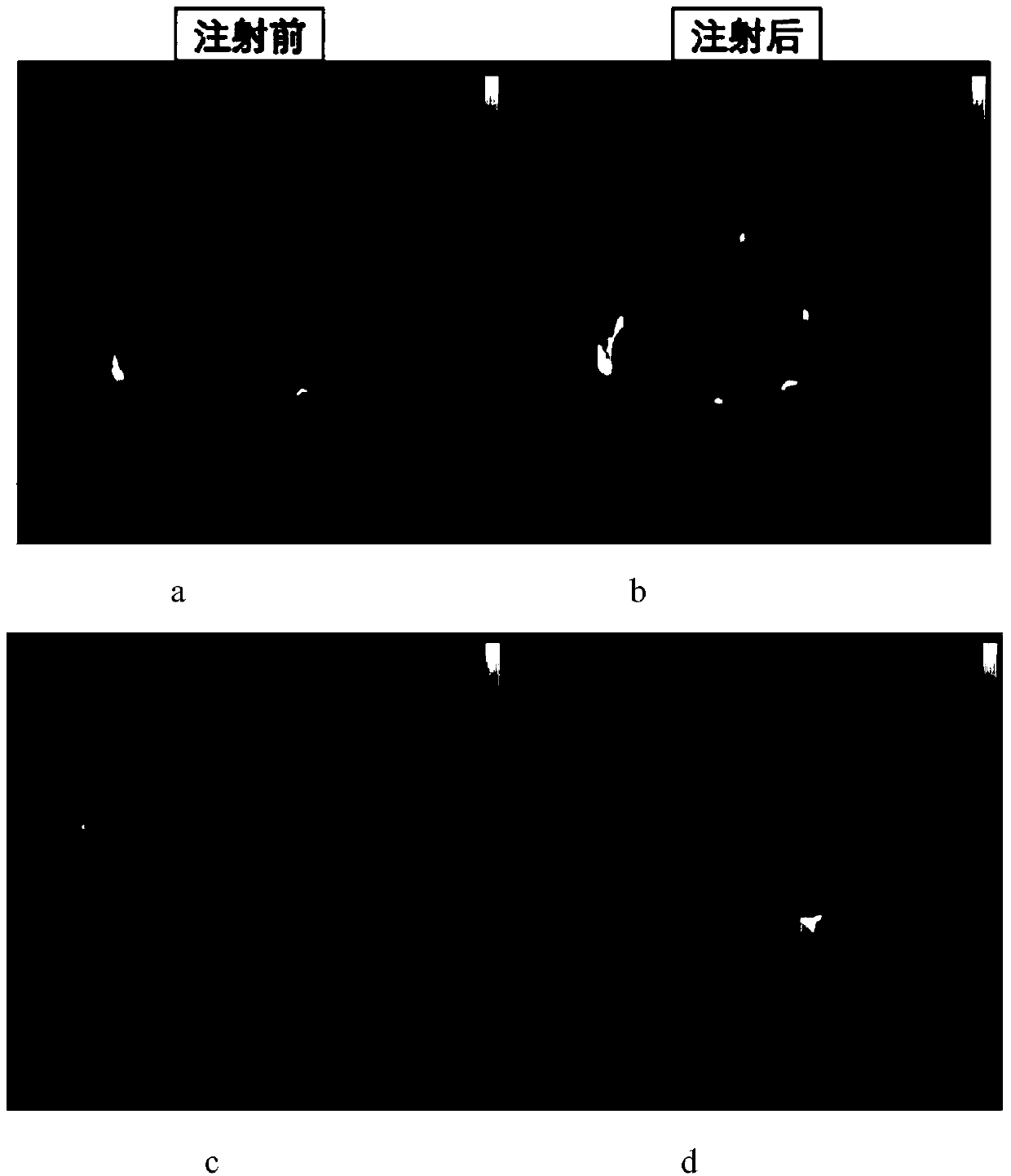
[0033] Nude mice were anesthetized with 5% isoflurane for photoacoustic tomography imaging of the brain and kidneys; then 20ml / kg of 20% fat emulsion was injected through the tail vein, and then photoacoustic imaging was performed on the brain and kidneys , to observe the enhancement effect of fat emulsion on photoacoustic imaging of animal tissue. Before the injection of fat emulsion, the signals of blood vessels in the brain, brain parenchyma and kidney tissue of nude mice were relatively weak, such as image 3 Shown in a and c. like image 3 As shown in middle b and d, after injecting fat emulsion, the photoacoustic signals of the brain blood vessels, brain parenchyma and kidney of nude mice were all significantly enhanced, indicating that the fat emulsion involved in blood circulation enhanced the imaging of the brain and kidney . Therefore, lipid emulsions can be used as contrast agents for photoacoustic imaging to enhance photoacoustic signals in animal tissues.


PUM


Abstract

The invention proposes the use of a fat emulsion as a photoacoustic imaging contrast agent, and belongs to the fields of biology and optics. The fat emulsion enhances the contrast ratio of a tissue photoacoustic signal and image. The fat emulsion is used for imaging animals or humans; for animal imaging, the fat emulsion is injected into animals through tail veins; and for human imaging, the fat emulsion is intravenously injected into the human body. The fat emulsion consists of soybean oil, lecithin and glycerin, and each 1000 ml of 20% fat emulsion contains 200 g of soybean oil, 12 g of lecithin, and 22 g of glycerin. The use of the fat emulsion as the photoacoustic imaging contrast agent can significantly enhance the contrast ratio of tissue photoacoustic imaging. In addition, the fat emulsion is often used as a supplement for intravenous nutrition in clinical practice. Compared with other photoacoustic imaging contrast agents, the fat emulsion is simple to manufacture, low in cost,and high in biosafety and has great potential for application as the imaging contrast agent.

Description

technical field [0001] The invention relates to the fields of biology and optics, in particular to the use of fat emulsion as a photoacoustic imaging contrast agent. Background technique [0002] Photoacoustic imaging is a new non-invasive and non-ionizing biomedical imaging method developed in recent years. Photoacoustic imaging combines the advantages of high selectivity in pure optical tissue imaging and deep penetration in pure ultrasonic tissue imaging to obtain high-resolution tissue images, and the photoacoustic signal transmits a large amount of information, which can provide morphological and functional information . Compared with other optical imaging techniques, such as optical coherence tomography, photoacoustic imaging can image deep tissues. However, some inherent chromophores in the body, such as hemoglobin, have weak absorption in the near-infrared band, resulting in weak contrast inside the tissue. Therefore, research on photoacoustic imaging contrast agen...

Claims


Application Information

Patent Timeline
IPC IPC(8): A61K49/22
CPCA61K49/22
Inventor 戚力张双阳刘嘉明理喜盼冯前进陈武凡
Owner SOUTHERN MEDICAL UNIVERSITY
Why Eureka
Try Eureka