Eureka AIR delivers breakthrough ideas for toughest innovation challenges, trusted by R&D personnel around the world.

Method for preserving umbilical cord membrane and preparing stem cells

A technology of stem cells and mesenchymal stem cells, which is applied in the field of isolating mesenchymal stem cells from the outermost amniotic membrane of the umbilical cord, can solve the problems of reduced number of cells, damage of mesenchymal stem cells, and long time required to achieve strong proliferation and differentiation potential , avoid fibroblasts and vascular endothelial cells, simple operation effect

Inactive Publication Date: 2019-04-19
北京弘润天源基因生物技术有限公司
View PDF10 Cites 3 Cited by
  • Summary
  • Abstract
  • Description
  • Claims
  • Application Information

AI Technical Summary

Problems solved by technology

Although the enzymatic digestion method can obtain cells in a short period of time, the cost is high, and the digestion time is difficult to grasp. The time required for digestion at room temperature is long, which is likely to cause damage to the isolated mesenchymal stem cells and lead to poor activity; the time required for digestion at 37°C Short, but the liquid is viscous, it is difficult to amplify the ideal number
The tissue block attachment method is easy to operate, has low requirements for experimental conditions, and is easy to master. The disadvantage of this method is that the acquisition of primary cells takes a long time, and the buoyancy of the liquid causes the tissue block to float. reduce

Method used


Image

  • Method for preserving umbilical cord membrane and preparing stem cells
  • Method for preserving umbilical cord membrane and preparing stem cells
  • Method for preserving umbilical cord membrane and preparing stem cells

Examples


Embodiment 1

[0035] Embodiment 1, methods for cryopreservation of umbilical cord membrane, recovery and separation and expansion of stem cells after recovery.

[0036] The method of processing and cryopreserving umbilical cord membranes includes the following steps.

[0037] (1) Collection of umbilical cord: under aseptic conditions in the delivery room, with the informed consent of the parturient, the umbilical cord of no less than 20 cm was intercepted immediately after the delivery of the fetus of the healthy parturient according to the obstetric routine ligation and umbilical cut. Rinse the umbilical cord with umbilical cord cleaning solution, then sterilize it with medical alcohol, put the umbilical cord in the umbilical cord preservation box and store it at a constant temperature of 2-8°C in a medical blood collection box. 4ml of peripheral blood was extracted from the mother for virus detection; 4ml of umbilical cord blood was extracted from the placenta for microbial detection.

...

Embodiment 2

[0047] Embodiment 2. Freezing storage of umbilical cord membrane, resuscitation and method for separation and expansion of stem cells after resuscitation

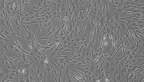
[0048] With reference to the method of Example 1, the revived umbilical cord membrane began to have adherent cells crawling out on the 7th day of culture, and the cell confluence reached 70% on the 13th day of culture. After the confluence reached more than 80%, the cells were digested with trypsin and subcultured into T25 culture flasks for culture. On the 18th day, the confluence reached 90%. After 2 passages, the cell purity reached more than 95%.

Embodiment 3

[0049] Embodiment 3. Freezing storage, resuscitation of umbilical cord membrane and separation and expansion method of stem cells after resuscitation

[0050] With reference to the method of Example 1, the revived umbilical cord membrane began to have adherent cells crawling out on the 8th day of culture, and the cell confluence reached 70% by the 14th day of culture. After the confluence reached more than 80%, the cells were trypsinized and subcultured into T25 culture flasks for culture. The confluence reached 90% on the 19th day. After 2 passages, the cell purity reached more than 95%.


PUM


Abstract

The invention relates to a method for preserving umbilical cord membrane and preparing stem cells. The umbilical cord membrane does not need to be attached to the wall during separation and culture, and the isolated mesenchymal stem cells have higher purity than that by an ordinary wartongel jelly tissue block adherence method. The warton jelly contains certain fibroblasts, which are very similarin characteristics to the mesenchymal stem cells and difficult to purify. The human amniotic membrane contains only two types of human amniotic epithelial cells and human amniotic mesenchymal stem cells. The epithelial cells have strong adherence ability and are easily removed during subculture, so that the obtained mesenchymal stem cells have higher purity. The method extracts the mesenchymal stem stem cells from the amniotic membrane layer covered by the outermost surface of the umbilical cord, and can effectively freeze and preserve the umbilical cord tissue for a long time, a frozen storage solution fully contacts the umbilical cord membrane, so that the activity of the cells on both sides of the umbilical cord membrane can be maximally maintained, resuscitation is convenient, especially separation of mesenchymal stem cells after resuscitation, and adherence treatment of umbilical cord tissue blocks cannot be required.

Description

technical field [0001] The invention belongs to the technical field of stem cell preparation, and in particular relates to a method for preparing human umbilical cord-derived mesenchymal stem cells, in particular to separating mesenchymal stem cells from the outermost amniotic membrane of the umbilical cord. Background technique [0002] Mesenchymal stem cells (MSCs) originate from the mesoderm in early development, and are a type of adult stem cells with high self-renewal ability and multilineage differentiation potential. MSCs have biological characteristics such as low immunogenicity, hematopoietic support, inflammatory chemotaxis, immune regulation, and nutritional support. They have been extensively studied in the fields of tissue injury, immune regulation, and regenerative medicine, and have broad application prospects. [0003] Mesenchymal stem cells have strong differentiation ability. In addition to differentiating into mesoderm cells such as fat, bone, cartilage, s...

Claims


Application Information

Patent Timeline
IPC IPC(8): C12N5/071C12N5/0775A01N1/02
CPCA01N1/0221A01N1/0284C12N5/0602C12N5/0665C12N2501/11C12N2501/115C12N2501/125C12N2509/00
Inventor 黄庆雷邓钺沈丽
Owner 北京弘润天源基因生物技术有限公司
Why Eureka
Eureka Blog
Learn More