
Biological sponge based on acellular dermal matrix and preparation method thereof

A decellularized dermis and decellularized technology, applied in the field of biosponge based on acellular dermal matrix and its preparation, can solve problems affecting clinical use, increasing costs, environmental pollution, etc., to ensure biological safety, enhance machinery, and reduce genotoxic effects

Active Publication Date: 2019-05-03
上海仁康科技有限公司
View PDF13 Cites 4 Cited by
  • Summary
  • Abstract
  • Description
  • Claims
  • Application Information

AI Technical Summary

Problems solved by technology

However, there are certain problems in these methods at present: most of the current cross-linking agents are reagents such as glutaraldehyde, and these reagents themselves will pollute the environment, and the follow-up treatment will increase the cost; the most important thing is that the residue of the reagents will cause severe cytotoxicity , which greatly affects clinical use

Method used


Image

  • Biological sponge based on acellular dermal matrix and preparation method thereof
  • Biological sponge based on acellular dermal matrix and preparation method thereof
  • Biological sponge based on acellular dermal matrix and preparation method thereof

Examples


Embodiment 1

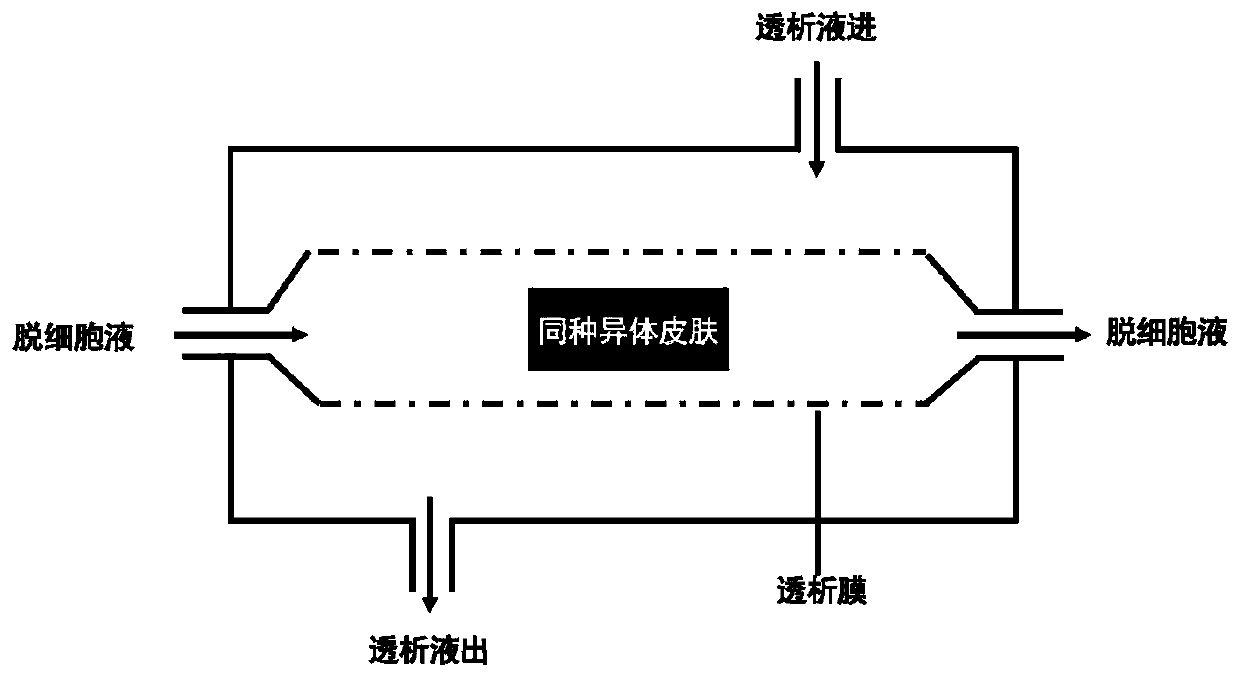
[0046] A preparation method of human-derived acellular dermal matrix, the specific steps are as follows:

[0047] (1) Preparation of acellular dermal matrix:

[0048] ①Take the stored human skin and remove the subcutaneous fatty components;

[0049] ②Step ①Clean and disinfect the skin with subcutaneous fatty components with povidone iodine and alcohol, and then soak and clean the skin repeatedly with hypotonic solution;

[0050] ③ Treat the skin washed in step ② with 2.5 mg / ml Dispase II solution overnight; discard the Dispase II solution, remove the epidermis, and then wash with normal saline to obtain the dermis;

[0051] ④Use the dermis obtained in step ③ with 2mol / L Ca 2+ NaCl solution for 18h, Ca 2+ The final concentration is 5mM / L; after discarding the sodium chloride solution, wash with normal saline;

[0052] ⑤ Place the dermis cleaned in step ④ into the prepared "decellularized solution", which contains the following components: 0.1%-0.5wt% trypsin, 0.5-5mM EDTA, ...

Embodiment 2

[0056] Internal structure of acellular dermal matrix

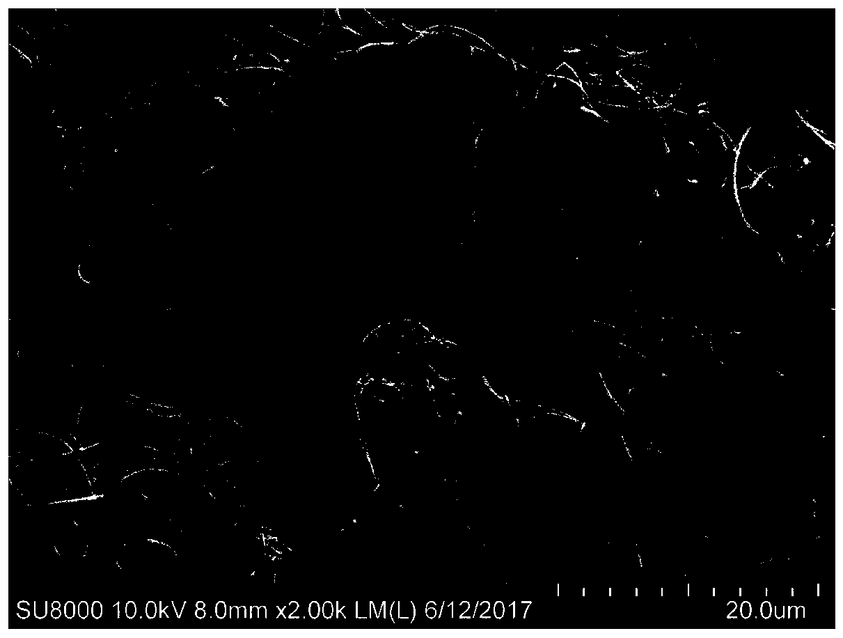
[0057] (1) Cut the acellular dermal matrix obtained above into a size of 1 cm × 1 cm, and paste it on a metal tray; the section of the acellular dermal matrix is ​​quenched with liquid nitrogen to make it hard and brittle, and then broken; the section faces Paste on the metal tray.

[0058] (2) After spraying gold in the sample preparation room of the electron microscope, it was put on the machine, observed and photographed by the scanning electron microscope, and the results were as follows: image 3 shown.

[0059] The results showed that there were no cell residues in the tissue, and the collagen structure was complete, similar to normal skin.

Embodiment 3

[0060] Example 3 HE staining of acellular dermal matrix

[0061] (1) Fix the normal skin tissue and the acellular dermal matrix in 4% paraformaldehyde fixative solution for 24 hours respectively;

[0062] (2) Alcohol dehydration, xylene transparency;

[0063] (3) Dip in wax for 30 minutes, embed, and make sagittal paraffin sections with a thickness of 5 μm;

[0064] (4) Gradient alcohol carries out dewaxing, dehydration, HE staining;

[0065] (5) Transparent, sealed, observed under a light microscope, the results of HE staining of normal skin tissue are as follows: Figure 4 As shown, the results of HE staining of acellular dermal matrix are as follows Figure 5 shown.

[0066] The results showed that there were no cell residues in the skin tissue.


PUM


Abstract

The invention discloses a biological sponge based on acellular dermal matrix and a preparation method thereof. The biological sponge is made of an acellular dermal matrix as a raw material, and Ca2<+>and fa-PEG are used for cross-linking. The biological sponge prepared by the invention has a porous structure, has pores which communicate with one another, and has a three-dimensional structure similar to human skin tissue, can enable body tissue cells to grow rapidly, has good plasticity, and can be prepared into different shapes as needed so as to meet the needs. The biological sponge of the invention is an advanced tissue engineering scaffold material, contains rich growth factors, has good biocompatibility, is more conducive to the growth of Sertoli cells, can not only be used for woundrepair, but also is an excellent tissue engineering skin scaffold material, and can be used for repairing skin defects and preparing tissue engineering skin.

Description

technical field [0001] The invention relates to the technical field of biomaterials, in particular to a biosponge based on decellularized dermal matrix and a preparation method thereof. Background technique [0002] Burns, wounds, diabetes, chronic ulcers, etc. often lead to skin defects. Pain, loss of physical function, and changes in appearance caused by refractory, chronic trauma, and tissue defects seriously affect the patient's freedom of movement and quality of life, and further lead to psychological and social problems. Skin transplantation is the most effective treatment for tissue defects and skin pigmentation diseases, but it is limited by sources in clinical practice, which affects the treatment process. Tissue-engineered skin is the most promising treatment method. The current tissue-engineered skin scaffold materials mainly include chemically synthesized materials such as polylactic acid (PLA), poly-left-lactic acid (PLLA), and the like. The material has stab...

Claims


Application Information

Patent Timeline
Patent Type & Authority Applications(China)
IPC IPC(8): A61L27/36A61L27/56A61L27/60
Inventor 董亚兵朱宁文朱迎春
Owner 上海仁康科技有限公司
Learn More