Recombinant cornea model for ocular surface irritation evaluation and preparation method thereof

An irritating, corneal technology, applied in biochemical equipment and methods, tissue regeneration, tissue culture, etc., can solve the problems of abnormal gene phenotype, limited to laboratory culture, unable to apply corneal model, etc., to improve the accuracy. Effect

Pending Publication Date: 2019-07-30
GUANGDONG BOXI BIO TECH CO LTD
View PDF1 Cites 0 Cited by
  • Summary
  • Abstract
  • Description
  • Claims
  • Application Information

AI Technical Summary

Problems solved by technology

Corneal epithelial cells are model tools for studying cell differentiation, signal transduction, and cell homeostasis signaling pathways in vitro. They can also be used to conduct three-dimensional corneal epithelial tissue models in vitro, but the limited sources of available corneal tissue, the short life cycle of corneal cells themselves and Rapid differentiation makes it difficult and time-consuming to culture corneal epithelial cells in vitro
Therefore, if the in vitro construction of three-dimensional corneal models using corneal epithelial cells does not solve the problem of the seed cells themselves, it will be limited to laboratory culture and cannot be industrialized. Many attempts to increase the in vitro culture cycle of corneal epithelial cells have been carried out. For example, virus transfection, telomerase reverse transcription gene transfection, etc., but these immortalized cell lines cannot be applied to the construction of corneal models in biology because of their abnormal gene phenotype and potential risk of tumorigenesis

Method used


Image

  • Recombinant cornea model for ocular surface irritation evaluation and preparation method thereof
  • Recombinant cornea model for ocular surface irritation evaluation and preparation method thereof
  • Recombinant cornea model for ocular surface irritation evaluation and preparation method thereof

Examples


Embodiment 1

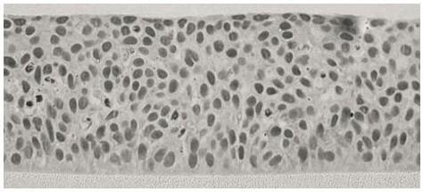
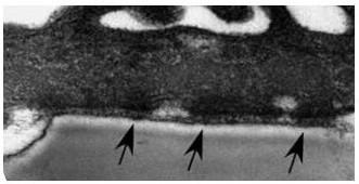
[0045] The in vitro reorganized corneal epithelial model in this implementation uses the corneal stroma of animals to simulate the growth microenvironment of limbal stem cells, and proliferates and differentiates during the in vitro induction period to form a corneal epithelial reorganized model with 4 layers of normal corneal epithelial cell layered structure. Hemidesmosomal proteins with a structure similar to tight junctions in natural tissues are formed between model basal cells and cellular compartments.

[0046] The preparation method of the above-mentioned in vitro recombinant corneal epithelial model consists of the following steps:

[0047] 1. Preparation of limbal stem cells

[0048] The eyeballs from the eye bank were soaked in saline containing 1% gentamicin for 30 minutes, and the cornea was cut along the limbus to obtain limbal tissue. Rinse the limbal tissue two or three times with D-Hank's solution. Cut the cleaned limbal tissue into 2 mm square tissue pieces...

Embodiment 2

[0056] The in vitro corneal epithelial model of this implementation adopts the corneal stroma layer of animals to simulate the growth microenvironment of limbal stem cells, and proliferates and differentiates during the in vitro induction period to form a corneal epithelial reorganization model with a layered structure of 6 layers of epithelial cells. Hemidesmosomal proteins with a structure similar to tight junctions in natural tissues are formed between model basal cells and cellular compartments.

[0057] Its preparation method is as follows:

[0058] 1. Preparation of limbal stem cells

[0059] This step is the same as in Example 1.

[0060] 2. In vitro culture and expansion of limbal stem cells

[0061] Take 10ml of the above-mentioned limbal stem cell cell suspension and 200mg of the corneal stromal microcarriers prepared in step 2 and inject into the rotary culture system with a capacity of 30ml, set the rotation speed at 30 rpm, change the medium with fresh DMEM ever...

Embodiment 3

[0067] The in vitro recombinant corneal epithelial model implemented in this implementation uses the corneal stroma layer of animals to simulate the growth microenvironment of corneal limbal stem cells, obtains large-scale expansion of corneal limbal stem cells, and uses corneal limbal stem cells and corneal epithelial cells to proliferate and differentiate in vitro. A model of corneal epithelial reorganization in layered cell hierarchy. Hemidesmosomal proteins with a structure similar to tight junctions in natural tissues are formed between model basal cells and cellular compartments.

[0068] 1. Preparation of limbal stem cells

[0069] This step is the same as in Example 1.

[0070] 2. In vitro culture and expansion of limbal stem cells

[0071] This step is the same as in Example 1.

[0072] 3. Construction of in vitro heavy corneal epithelial model

[0073] 3.1 Resuspend the limbal stem cells obtained in step 2 with culture medium SK1, inoculate in a cell chamber with...


PUM


Abstract

The invention relates to a recombinant cornea model for ocular surface irritation evaluation and a preparation method thereof, in particular to the field of in-vitro organ culture and tissue regeneration. From the perspective of seed cells, by constructing the three-dimensional corneal epithelium model through in-vitro enrichment and corneal limbal stem cell amplification, the in-vitro in-vivo consistency of eye irritatwion evaluation is ensured with the seed cells as a cornea family, the problem about industrial large-scale production is also solved, and the promotion of ocular surface irritation in-vitro substitutive experiments is facilitated.

Description

technical field [0001] A recombined corneal model and a preparation method thereof for evaluation of ocular surface irritation, specifically relate to the fields of in vitro organ culture and tissue regeneration. Background technique [0002] In recent years, with the development of in vitro organ culture technology, the 3D recombinant corneal model based on in vitro cell culture has remarkable efficacy in clinically replacing damaged corneal structures and repairing corneal damage. However, because its clinical application is mainly to replace corneal damaged tissue for transplantation, it is mostly produced in the form of an open sheet, and it is a three-dimensional space complex formed by combining biological materials and different types of seed cells in vitro, so the structure of the entire corneal tissue and Permeability has changed, and the evaluation of chemical eye irritation needs to imitate the penetration process of chemicals on the ocular surface, and it is requ...

Claims


Application Information

Patent Timeline
IPC IPC(8): C12Q1/02C12N5/074A61L27/38A61L27/50A61F2/14
CPCG01N33/5088C12N5/0621C12N5/0623A61L27/3804A61L27/3839A61L27/3895A61L27/50A61F2/142C12N2531/00A61L2430/16A61L2430/40
Inventor 李潇卢永波邓志宏
Owner GUANGDONG BOXI BIO TECH CO LTD
Why Eureka
Try Eureka