Method for in-situ observation of cell connection between monolayer cells by using transmission electron microscope

A technique of single-layer cells and transmission electron microscopy, which is applied to material analysis, measuring devices, and instruments using wave/particle radiation. It can solve problems such as complicated steps, long time-consuming, and expensive consumables, and achieve stable methods and reduced damage. Effect

Active Publication Date: 2019-08-02
曾琪琪
View PDF2 Cites 3 Cited by
  • Summary
  • Abstract
  • Description
  • Claims
  • Application Information

AI Technical Summary

Problems solved by technology

This method is mainly used to observe the structure of Schwann cells and Schwann cell dorsal root ganglia in situ. Its shortcoming is that the glass slide is used as the cell growth carrier, and the resin specimen needs to be corroded by hydrofluoric acid. , hydrofluoric acid can have a certain impact on the cell specimen on the resin surface or on the cell connection th

Method used


Image

  • Method for in-situ observation of cell connection between monolayer cells by using transmission electron microscope
  • Method for in-situ observation of cell connection between monolayer cells by using transmission electron microscope
  • Method for in-situ observation of cell connection between monolayer cells by using transmission electron microscope

Examples


Embodiment 1

[0045] Material preparation: cells and their growth carriers: human pancreatic ductal epithelial cell line (HPDE6C7), Epon-812 epoxy resin sheet of 15mm×20mm×0.5mm.

[0046]Cell preparation: (1) HPDE6C7 cells are growing well, covering about 90% of the T25 culture flask; (2) Put the Epon-812 epoxy resin slices irradiated by the ultraviolet lamp on both sides into the culture plate; (3) 0.25% pancreatic Digest the cells with enzymes, centrifuge, resuspend, and count the cells. Add the cells evenly to a six-well plate with resin slices at a cell density of 1×10^6 / ml / well, and place them in a 37°C, 5% CO 2的 Incubator cultivation.

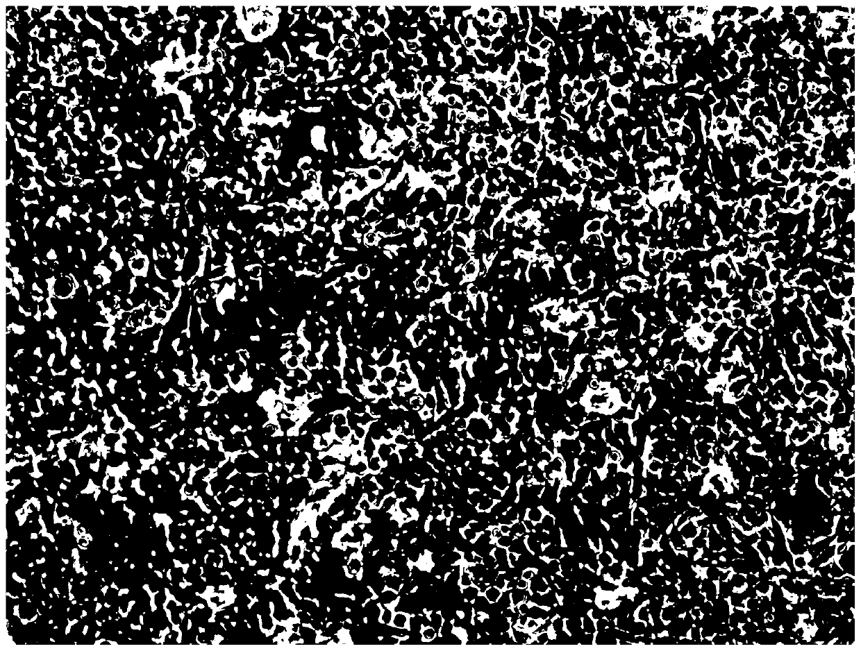
[0047] Specimen fixation, dehydration, and soaking: (1) After the cells cover more than 90% of the resin sheet, add 3% glutaraldehyde fixative solution and place it at 4°C for 12-16h; figure 1 It shows that HPDE6C7 cells are well cultivated in Epon-812 epoxy resin sheet; (2) 0.1mol / L phosphate buffered saline (PBS) washes 3 times, each time 8min; (3) ...

Embodiment 2

[0052] (1) Cell preparation: HPDE6C7 cells are growing well, covering about 90% of the T25 culture flask; (2) Put the Epon-812 epoxy resin slices irradiated by the ultraviolet lamp on both sides into the culture plate; (3) 0.25% pancreatic Digest the cells with enzymes, centrifuge, resuspend, and count the cells. Add the cells evenly to a six-well plate with resin slices at a cell density of 1×10^6 / ml / well, and place them in a 37°C, 5% CO 2的 incubator cultivation;

[0053] (2) Specimen fixation, dehydration, soaking:

[0054] a. Specimen fixation: add the cells cultivated in step (1) to glutaraldehyde fixative solution, place at 4°C for 12 hours, wash with 0.1mol / L phosphate buffer solution for 3 times, each time for 8 minutes; then use 1% osmium After acid fixation for 1 hour, wash with 0.1mol / L phosphate buffer 3 times, each time for 8 minutes;

[0055] b. Ethanol step-by-step dehydration: 50% ethanol, 70% ethanol, 90% ethanol dehydration once respectively, 100% ethanol de...


PUM

PropertyMeasurementUnit
Thicknessaaaaaaaaaa
Thicknessaaaaaaaaaa

Abstract

The invention relates to the technical field of cell observation, and particularly relates to a method for in-situ observation of cell connection between monolayer cells by using a transmission electron microscope. The method comprises the following steps: (1) cell preparation: putting sterilized epoxy resin sheets into a culture plate, centrifuging and resuspending the single-layer cells, uniformly adding the single-layer cells into a substrate with the epoxy resin sheets, and putting the substrate into an incubator for culture; (2) specimen fixing, dehydrating and soaking; (3) area marking,block repairing and pre-embedding; (4) pre-embed block repairing and re-embedding; and (5) slicing, dyeing and electron microscope observation. Through the steps, the original appearance of the monolayer cells can be restored to the greatest extent in situ. Compared with the traditional method, the method has the advantages that the damage to cell connection is reduced; compared with a foreign in-situ embedding method, the resin serving as a carrier for cell growth is simpler and easier to implement compared with a glass slide, and the pre-embedding and re-embedding method is more stable and effective.

Description

technical field [0001] The invention relates to the technical field of cell observation, in particular to a method for in-situ observation of cell junctions between monolayer cells with a transmission electron microscope. Background technique [0002] Cell junction is the connection structure between cells, which is formed by the specialization of the local area of ​​the plasma membrane, and structurally includes the membrane specialized part, the cytoplasmic part under the plasma membrane and the intercellular part outside the plasma membrane. In multicellular organisms, biological networks of cell connections exist widely, and adjacent cells communicate with each other through plasma membranes, forming a complex biological communication system. TEM enables unique and valuable studies of intracellular and extracellular material extracted from tissues in situ, cultured cells, and suspension cells. [0003] The sample preparation method for traditional adherent cultured cell...

Claims


Application Information

Patent Timeline
IPC IPC(8): G01N23/2251G01N23/2202
CPCG01N23/2251G01N23/2202G01N2223/07G01N2223/102G01N2223/612
Inventor 曾琪琪唐国都杨慧莹梁志海
Owner 曾琪琪
Why Eureka
Try Eureka