Amnion contact lens, preparation method and application

A contact lens and amniotic membrane technology, applied in the field of medicine, can solve the problems of easy damage to amniotic membrane and cornea, scarring of corneal tissue, insufficient drug loading, etc.

Pending Publication Date: 2019-08-16
GUANGDONG PROPHET BIOTECHNOLOGY CO LTD
View PDF7 Cites 6 Cited by
  • Summary
  • Abstract
  • Description
  • Claims
  • Application Information

AI Technical Summary

Problems solved by technology

[0007] The embodiment of the present invention provides an amniotic contact lens, a preparation method and its application, which are used to solve the problem of insufficient drug loading in existing contact lenses, and the fact that organic solvents are easy to damage the amniotic membrane and cornea, resulting in scarring of corneal tissue formed problem

Method used


Image

  • Amnion contact lens, preparation method and application
  • Amnion contact lens, preparation method and application
  • Amnion contact lens, preparation method and application

Examples


Embodiment 1

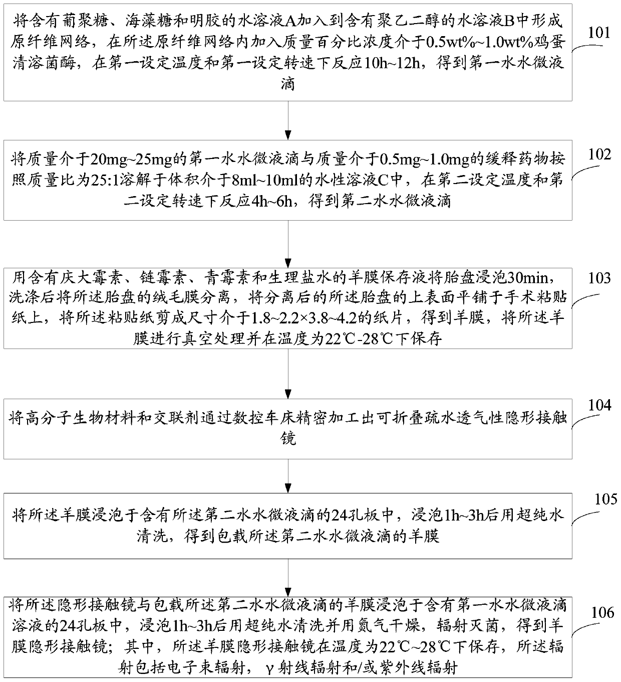
[0052] Step 101, adding the aqueous solution A containing dextran, trehalose and gelatin to the aqueous solution B containing polyethylene glycol to form a fibril network, and adding a concentration of 0.5 wt% to 1.0 wt% in the fibril network Wt% egg white lysozyme is reacted at the first set temperature and the first set speed for 10h to 12h to obtain the first water micro-droplet;

[0053] Step 102, dissolving the first water micro-droplet with a mass of 20mg-25mg and the slow-release drug with a mass of 0.5mg-1.0mg in an aqueous solution C with a volume of 8ml-10ml according to a mass ratio of 25:1 , reacting at the second set temperature and the second set speed for 4h to 6h to obtain the second water droplets;

[0054] Step 103, soak the placenta in amniotic membrane preservation solution containing gentamicin, streptomycin, penicillin and normal saline for 30 minutes, separate the chorion of the placenta after washing, and tile the upper surface of the separated placenta...

Embodiment 2

[0077] Step 201, adding 1.5wt%, 0.5wt%, and 0.5wt% dextran, trehalose and gelatin solutions to a 6.5wt% polyethylene glycol solution to form a fibril network , and then add 1wt% egg white lysozyme, and react for 10 hours at a temperature of 60° C. with a rotation speed of 100 rpm, thereby forming first water droplets with a diameter of 5 nm.

[0078] Step 202, dissolving 25 mg of the first water micro-droplet and sustained-release drug in a mass ratio of 25:1 in 10 ml of an aqueous solution, reacting for 5 hours at a temperature of 4°C and a rotation speed of 250 rpm, and finally The second water micro-droplets are obtained.

[0079] Step 203, soak the placenta with amniotic membrane preservation solution containing penicillin, streptomycin, penicillin and normal saline for 30 minutes, wash several times until the blood stains on the placenta are washed away, then bluntly separate the amniotic membrane from the chorion of the placenta, and put on Spread the surface on the sur...

Embodiment 3

[0086] Step 301 Preparation of the first water micro-droplets: adding dextran, trehalose, and gelatin with mass percent concentrations of 1.0wt%, 0.1wt%, and 0.1wt% respectively to polyethylene glycol containing 5.5wt% mass percent concentration A fibril network is formed in a solution of diol, and then a percentage concentration of 0.5 wt% egg white lysozyme is added, and the temperature is 65° C. and the rotation speed is 200 rpm, and the reaction is carried out for 12 hours, and the first fiber with a diameter of 5 nm can be prepared. Water water micro-droplets.

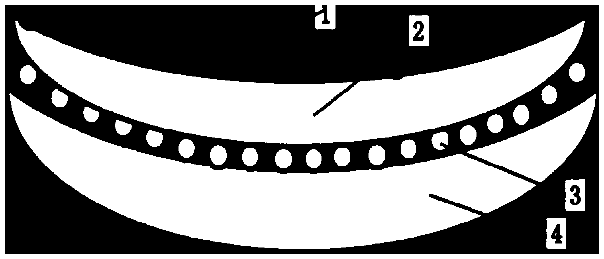
[0087] In order to further illustrate that the contact lens and the amnion in the amniotic contact lens provided by the embodiments of the present invention have good adhesion, preferably, the contact lens prepared by Example 1 or Example 2 and the entrapped The amniotic membrane of the second water micro-droplet is soaked in the 24-well plate containing the first water micro-droplet provided in Example 3, after s...


PUM


Abstract

The invention relates to the field of medicine, discloses an amnion contact lens, a preparation method and application and aims to solve problems that existing contact lenses are insufficient in drugloading capacity and organic solvents are prone to injuring the amnion and the cornea and causing cornea tissue scarring. The amnion contact lens comprises a contact lens body, the amnion, first water-water microdroplets and second water-water microdroplets. The first water-water microdroplets are arranged on the inner surface of an optical area of the contact lens body; the amnion is adhered on the inner surface of an optical area of the contact lens body through the first water-water microdroplets; the second water-water microdroplets are adhered to the inner surface of the amnion. Controlled-release medicines are wrapped in the second water-water microdroplets, and the contact lens has advantages of foldability, hydrophobicity and breathability.

Description

technical field [0001] The invention relates to the technical field of medicine, in particular to an amniotic membrane contact lens, a preparation method and an application thereof. Background technique [0002] The cornea is at the forefront of the eye and is generally the most vulnerable to trauma. After the cornea is injured, the surrounding epithelial cells will continue to provide the cells needed by the damaged site until it is fully restored. But it is easy to form scar tissue, resulting in visual impairment. Blindness caused by corneal injury is caused by scar tissue. Common causes of intraocular trauma include: 1. Trauma: corneal perforation injury, explosion injury, chemical burn, thermal burn, etc.; 2. Infectious corneal injury: including bacteria, fungi, Corneal diseases caused by viruses; 3. Surgical trauma: Refractive surgery, cataract surgery, etc. The biggest obstacle to the development of surgery is the postoperative scar formation in the hand and eye, and...

Claims


Application Information

Patent Timeline
Patent Type & Authority Applications(China)
IPC IPC(8): A61F9/00A61K9/00A61K45/00A61K47/46A61P27/02A61P27/06A61P29/00
CPCA61F9/0017A61K9/0051A61K47/46A61K45/00A61K9/0087A61P27/02A61P29/00A61P27/06
Inventor 周辉
Owner GUANGDONG PROPHET BIOTECHNOLOGY CO LTD
Why Eureka
Try Eureka