Cryoprotectant for umbilical cord mesenchymal stem cells and application of cryoprotectant

A technology of mesenchymal stem cells and cryopreservation solution, which is applied in the field of umbilical cord mesenchymal stem cell cryopreservation solution, can solve the problems of clinical application limitations of mesenchymal stem cells, complex components of fetal bovine serum, side effects of antigens and antibodies, etc., and optimize the preparation volume and component concentration, the effect of good freezing effect

Inactive Publication Date: 2019-09-06
汪文
View PDF4 Cites 5 Cited by
  • Summary
  • Abstract
  • Description
  • Claims
  • Application Information

AI Technical Summary

Problems solved by technology

[0004] At present, the traditional cell cryopreservation solution contains basal medium, dimethyl sulfoxide, fetal bovine serum, etc., but the composition of fetal bovine serum is complex and contains a large number of alloantigens, which is difficult to clean in the subsequent recovery and culture process, and it is returned to the The human body causes antigen-antibody side effects, so it is currently

Method used


Image

  • Cryoprotectant for umbilical cord mesenchymal stem cells and application of cryoprotectant
  • Cryoprotectant for umbilical cord mesenchymal stem cells and application of cryoprotectant
  • Cryoprotectant for umbilical cord mesenchymal stem cells and application of cryoprotectant

Examples


Embodiment 1

[0063] A cryopreservation method for umbilical cord mesenchymal stem cells is provided, comprising the steps of:

[0064] (1) Wash the umbilical cord tissue (remove blood cells): add an equal amount of normal saline to the centrifuge tube containing the umbilical cord, tighten the cap, shake for 3 minutes to fully wash the umbilical cord tissue, then stand still for 3-5 minutes to separate the different phases, and suck off the lower layer Water phase; repeat the above operation three times until the lower layer is relatively clear.

[0065] (2) Ultrasonic microwave pulverization: After aspirating and discarding the normal saline, add preheated DMEM culture solution equal to the volume of the umbilical cord, place in a constant temperature microwave pulverizer, and ultrasonically pulverize at 37°C.

[0066] (3) Collect the precipitate: After digestion, centrifuge at 2000rpm for 10min, discard the digested umbilical cord in the upper layer, collect the bottom layer of the two t...

Embodiment 2

[0073] Identification of umbilical cord stem cells after cryopreservation

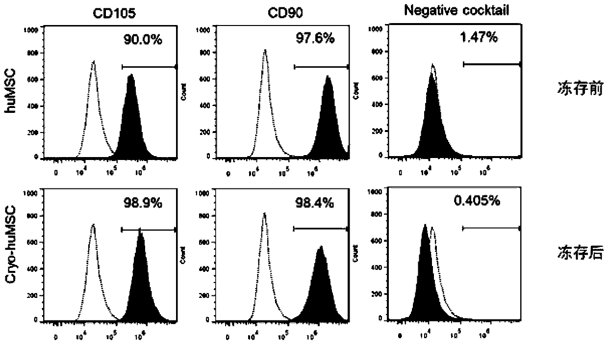
[0074] The P2 umbilical cord stem cells cultured in Example 1 were centrifuged and resuspended; after cell counting, the cell concentration was adjusted to 1×10 8 / L, react with human anti-CD105, CD90, CD34 and CD45 mixture (Cocktail) monoclonal antibody for 30min at room temperature, resuspend the cells with PBS, and detect with flow cytometry ( figure 2 ).

[0075] See the test results figure 2 It can be seen that the analysis of cell surface antigen marker expression of umbilical cord stem cells by flow cytometry before and after cryopreservation shows that the phenotype of umbilical cord mesenchymal stem cells has no obvious change after cryopreservation in the modified cryopreservation medium.

Embodiment 3

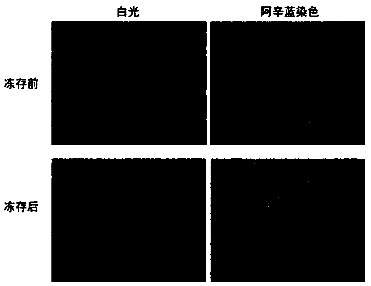
[0077] Chondrogenic ability test of umbilical cord stem cells after cryopreservation

[0078] The cells frozen in Example 1 were used as the group of the present invention, and the chondrogenic ability test was carried out. After 3-4 weeks of in vitro differentiation towards cartilage, see image 3 It can be seen that Alcian blue staining shows that the umbilical cord mesenchymal stem cells cryopreserved in Example 1 and the umbilical cord mesenchymal stem cells not cryopreserved have the same ability to differentiate into cartilage in vitro, and there is no obvious difference in ability.


PUM

PropertyMeasurementUnit
Volumeaaaaaaaaaa
Volumeaaaaaaaaaa

Abstract

The invention discloses a cryoprotectant for umbilical cord mesenchymal stem cells and application of the cryoprotectant. The cryoprotectant comprises a human serum albumin solution, a dimethyl sulfoxide solution, trophic factors and a pharmaceutically-acceptable carrier, and the trophic factors are one or more of a vascular endothelial cell growth factor VEGF, a transforming growth factor TGF, interleukin 6IL-6, a fiber bud cell growth factor bFGF and a hepatocyte growth factor HGF. The invention discloses the application of the cryoprotectant for the umbilical cord mesenchymal stem cells incryopreservation of the umbilical cord mesenchymal stem cells. Through the above mode, according to the application, the specific cryoprotectant is adopted to cryopreserve the umbilical cord mesenchymal stem cells, allows the cryopreserved umbilical cord mesenchymal stem cells to keep activity and differentiation stemness, and has a good cryopreservation effect.

Description

technical field [0001] The invention relates to the technical field of cell cryopreservation, in particular to a cryopreservation solution for umbilical cord mesenchymal stem cells and its application. Background technique [0002] Cell cryopreservation is a technique in which cells are placed in a low temperature environment to reduce cell metabolism for long-term storage. Cell cryopreservation is one of the main methods of cell preservation, which plays a role in cell preservation. [0003] Cell cryopreservation is the use of cryopreservation technology to store cells in liquid nitrogen at -196°C at low temperature, which can temporarily remove the cells from the growth state and preserve their cell characteristics, so that the cells can be revived for clinical application when needed. Properly preserving a certain amount of cells can prevent the cells from being lost due to contamination of the cells being cultured or other accidents, and play a role in cell preservation...

Claims


Application Information

Patent Timeline
IPC IPC(8): A01N1/02
CPCA01N1/021A01N1/0226
Inventor 汪文
Owner 汪文
Why Eureka
Try Eureka