Medical image analysis method based on computer vision

A computer vision and medical image technology, applied in the field of image processing, can solve the problems of incompatible data format, provide resources, and inconvenient data transmission, so as to improve the utilization rate and circulation, reduce difficulty and cost, and realize data sharing. Effect

Inactive Publication Date: 2019-09-06
JIANGSU UNIV OF SCI & TECH IND TECH RES INST OF ZHANGJIAGANG
View PDF0 Cites 4 Cited by
  • Summary
  • Abstract
  • Description
  • Claims
  • Application Information

AI Technical Summary

Problems solved by technology

Radiologists can find and diagnose the disease by observing images, but this diagnosis method has the following main shortcomings: (1) The contrast of X-ray images (including CT) is very low, and it is easy to misdiagnose (2) Nuclear isotope images and ultrasound images Rough and low resolution; (3) It is difficult and inaccurate to construct a 3D structure from a set of 2D images
[0005] The current digital management of medical images is difficult and costly, and the management is not standardized, so it is not suitable for image query and retrieval; storage requires film, which is inconvenient for data transmission, and data is easily damaged, deteriorated, and lost; it is not compatible with current large The data format of some medical imaging equipment manages various medical images, and cannot make full use of the Internet to realize data sharing, remote diagnosis, and consultation in multiple places. It cannot provide resources for medical teaching, and the utilization rate and circulation of data are poor.

Method used


Image

  • Medical image analysis method based on computer vision

Examples


Embodiment Construction

[0016] Exemplary embodiments of the present disclosure will be described in more detail below with reference to the accompanying drawings. Although exemplary embodiments of the present disclosure are shown in the drawings, it should be understood that the present disclosure may be embodied in various forms and should not be limited by the embodiments set forth herein. Rather, these embodiments are provided for more thorough understanding of the present disclosure and to fully convey the scope of the present disclosure to those skilled in the art.

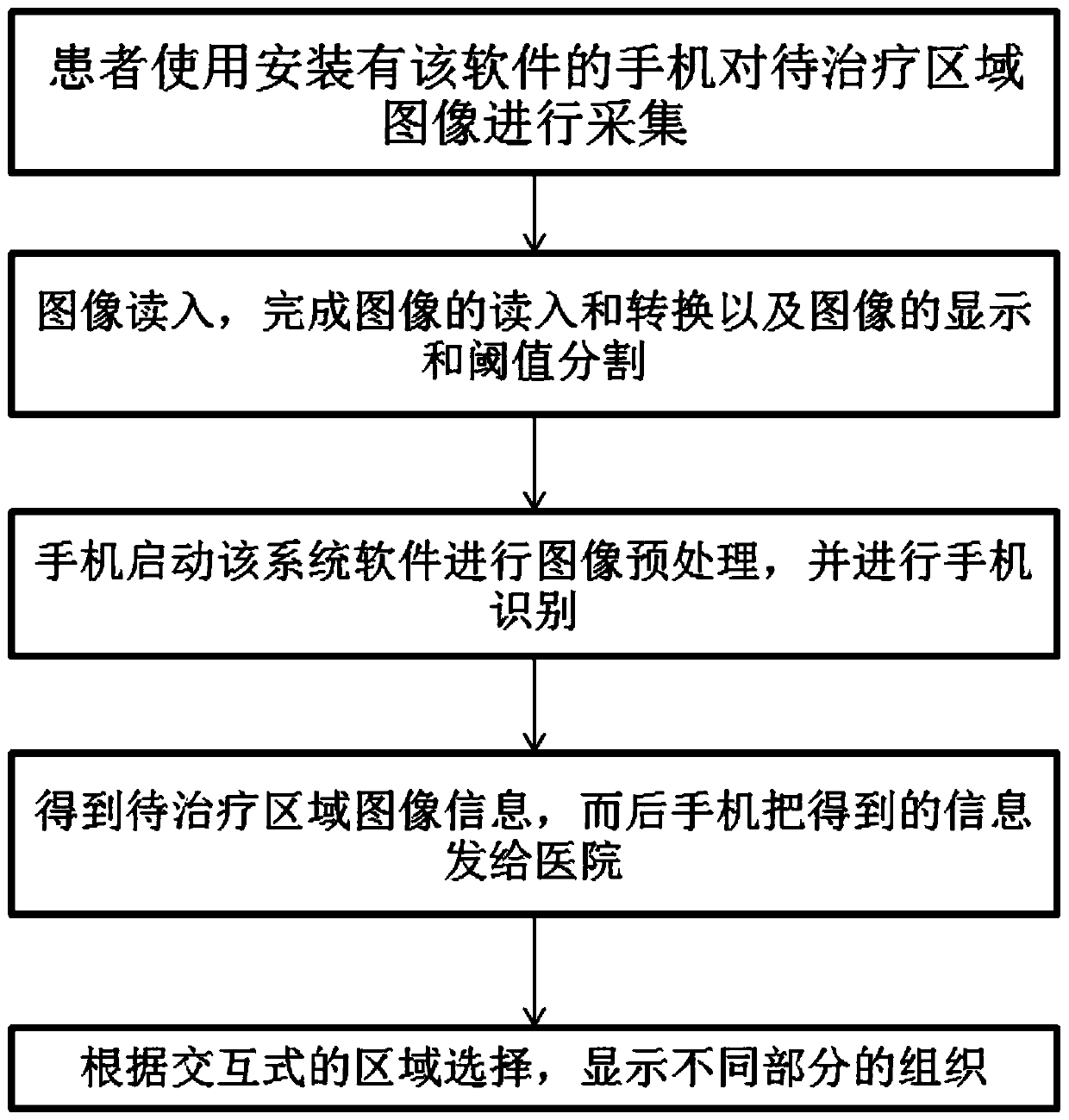
[0017] Refer to attached figure 1 It is a working flowchart of a computer vision-based medical image analysis method involved in the present invention.

[0018] The present invention claims a medical image analysis method based on computer vision, which is characterized in that it includes:

[0019] The patient uses the mobile phone installed with the software to collect images of the area to be treated: if the collected image is ...


PUM


Abstract

The invention requests to protect a medical image analysis method based on computer vision to complete preprocessing work of an input medical image sequence, the preprocessing work mainly comprises gray scale transformation, smoothing and noise elimination, and image segmentation of a preprocessed medical image is achieved. Digital management of medical images is realized, the management difficulty and cost are reduced, the management is more standard, and the method is more suitable for query and retrieval of the images; the image is stored in a digital form, the information of the image is completely reserved, the window position and the window width can be changed, and some image processing technologies can be used for processing; a magnetic disk is used for image storage, no film is needed any more, data transmission is facilitated, and the data is not prone to damage, deterioration and loss; and the DICOM standard is adopted, so that data formats of most of current medical image equipment can be compatible, and the utilization rate and the liquidity of data are greatly improved.

Description

Background technique [0001] The present invention relates to the technical field of image processing, in particular to a computer vision-based medical image analysis method, which is suitable for target tracking, target recognition, video surveillance, video conferencing, biomedicine, information security, remote sensing and telemetry, aerospace or video multimedia Image matching. Background technique [0002] Since the 1970s, with the emergence and application of medical imaging equipment such as computerized tomography and nuclear magnetic resonance imaging, people can obtain two-dimensional digital tomographic image sequences of the human body and its internal organs. Radiologists can find and diagnose the disease by observing images, but this diagnosis method has the following main shortcomings: (1) The contrast of X-ray images (including CT) is very low, and it is easy to misdiagnose (2) Nuclear isotope images and ultrasound images It is relatively rough and the reso...

Claims


Application Information

Patent Timeline
Patent Type & Authority Applications(China)
IPC IPC(8): G06T7/136G06T5/40G06T5/00
CPCG06T5/002G06T5/40G06T2207/20032G06T2207/20064G06T7/136
Inventor 周塔陶献高尚徐正涛张鑫于静薛伟郭凌王思琦张宁
Owner JIANGSU UNIV OF SCI & TECH IND TECH RES INST OF ZHANGJIAGANG
Why Eureka
Try Eureka