Enhanced anti-tumor fusion protein, preparation method and use thereof

A fusion protein and anti-tumor technology, which is applied in the field of fusion protein and preparation, to achieve obvious killing effect

Active Publication Date: 2019-10-08
孙嘉琳 +1
View PDF8 Cites 2 Cited by
  • Summary
  • Abstract
  • Description
  • Claims
  • Application Information

AI Technical Summary

Problems solved by technology

[0007] At present, there is no report on the combination of various inducing or stimulating T cell molecules for tumor therapy

Method used


Image

  • Enhanced anti-tumor fusion protein, preparation method and use thereof
  • Enhanced anti-tumor fusion protein, preparation method and use thereof
  • Enhanced anti-tumor fusion protein, preparation method and use thereof

Examples


Embodiment 1

[0036] Example 1 Construction of vectors expressing various proteins

[0037] The expression plasmid adopted is pET22b (Novagen Company), which contains various restriction enzyme cutting sites (BamHI-EcoRI-SacI-SalI-HindIII-NotI-XhoI) for the insertion of DNA fragments encoding foreign proteins, and outside expression The C-terminus of the source protein has a His-tag (containing 6 His) for protein purification.

[0038] DNA fragments were synthesized by TAKARA Company and Shanghai Sangon Bioengineering Company.

[0039] The nucleic acid sequence of synthetic B7.1 is shown by SEQ ID No.1;

[0040] The nucleic acid sequence of synthetic B7.2 is shown by SEQ ID No.3;

[0041] The nucleic acid sequence of the synthetic SEA is shown in SEQ ID No.5;

[0042] The nucleic acid sequence of the synthetic B7.1-B7.2 fusion protein is shown in SEQ ID No.7, and there is a nucleic acid fragment (gtcgacaagctttccggcggaggtggc) (shown in SEQ ID No.15) of a connecting peptide between B7.1 an...

Embodiment 2

[0057] Example 2 Expression, denaturation and renaturation and purification of various proteins

[0058] Various expression plasmids pET22b-B7.1, pET22b-B7.2, pET22b-SEA, pET22b-B7.1-B7.2, pET22b-B7.1-SEA, pET22b-B7.2-SEA and pET22b-B7 .1-B7.2-SEA were transformed into Escherichia coli BL21(DE3) by electroporation, and the positive bacteria were screened by antibiotic Amp (Ampicillin). Next, the process of expression, denaturation, renaturation and purification of various proteins is roughly the same, and the operation is as follows:

[0059] Escherichia coli BL21(DE3) containing the expression plasmid was first cultured at 37°C on a large scale, and then IPTG (Isopropylthio-β-D-galactoside) was added to make the concentration reach 1mM and cultured at 30°C overnight to induce protein expression. On the second day, the culture medium was centrifuged and the bacteria were collected, the cell wall was broken by ultrasonic method, and the inclusion body precipitate was collected...

Embodiment 3

[0060] Embodiment 3 tumor suppression experiment

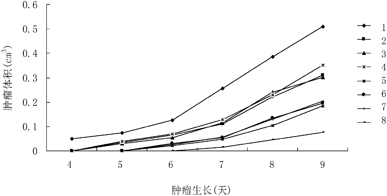
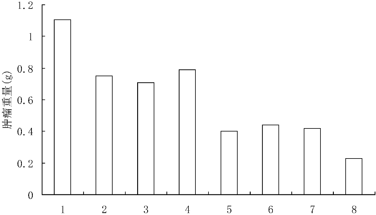
[0061] Select male ICR mice, 4-5 weeks old, 18-22g, and divide them into 8 groups randomly, 30 mice in each group. Mouse sarcoma cells S180 were cultured from mouse ascites, and 5x10 5 Mouse sarcoma cells S180 were inoculated in the right armpit of the mice, and then B7.1, B7.2, SEA, B7.1-B7.2, B7.1- SEA, B7.2-SEA and B7.1-B7.2-SEA protein solution 0.2ml (500pmol) / only, and control group injected equal amount of normal saline (0.9%NaCl), the 9th day put to death mice, observed small The tumor growth of the mice and the weight of the tumors after the mice were sacrificed were measured.

[0062] figure 1 Indicates that various proteins inhibit tumor growth, the horizontal axis in the figure represents the growth days of tumor-bearing mice, and the vertical axis represents the tumor volume (cm 3 ), the number of the right curve in the figure indicates the grouping of the experimental mice, 1 is the normal saline group, 2 is t...


PUM


Abstract

The invention discloses an enhanced anti-tumor fusion protein, a preparation method and use thereof. The fusion protein includes at least two of a costimulatory molecule B7.1 that induces and activates T cells, a costimulatory molecule B7.2 that induces and activates the T cells, and a superantigen Staphylococcus aureus enterotoxin A. The fusion protein of the present invention constructs at leasttwo of B7.1, B7.2 and SEA into the fusion protein, and exerts the superimposed amplification effect. The biological activity is far more than that of a single source protein, the T cells are inducedand activated to kill tumors, the T cells secrets cytokines such as tumor necrosis factor alpha (TNF-alpha) and granzymes and induces the cancer cells as target cells to produce Fas and the like to cause cancer cells to apoptosis, The experiments have shown that each of the fusion proteins of the present invention has the significant killing effect on various cancers and solid tumors.

Description

technical field [0001] The present invention relates to a fusion protein and its preparation method and application, in particular to a fusion protein composed of co-stimulatory molecules (Costimulatory molecules) that induce and activate T cells and superantigen Staphylococcus aureus enterotoxin that can cause anti-cancer immune response The fusion proteins of , disclosed their structure (protein sequence), preparation method and anti-cancer biological activity. Background technique [0002] The effective activation of T cells requires the participation of two signals, that is, the combination of the antigen peptide-MHC complex with TCR-CD3 on T cells provides the first signal, and the second signal mediated by costimulatory molecules. The most basic co-stimulatory signal is provided by the B7.1 (CD80) and B7.2 (CD86) molecules expressed on antigen-presenting cells and the corresponding receptors (CD28 and CTLA-4) expressed on T cells (Amj Respir Crit Care Med , 162, 5164-...

Claims


Application Information

Patent Timeline
Patent Type & Authority Applications(China)
IPC IPC(8): C07K19/00C12N15/62A61K38/17A61K38/16A61P35/00
CPCC07K14/31C07K14/70532A61P35/00C07K2319/00C07K2319/55A61K38/00
Inventor 孙嘉琳
Owner 孙嘉琳
Why Eureka
Try Eureka