Method for differentiating 5-methylcytosine and 5-hydroxymethylcytosine in DNA

A technology for hydroxymethylated cytosine and methylated cytosine, which is applied in the field of distinguishing 5-methylated cytosine and 5-hydroxymethylated cytosine in DNA, and can solve the problem of large sequence preference and low transformation efficiency and other problems, to achieve the effect of improving timeliness, low damage rate, and increasing stability

Pending Publication Date: 2019-12-03
WUHAN BIORUN BIO TECH
View PDF5 Cites 0 Cited by
  • Summary
  • Abstract
  • Description
  • Claims
  • Application Information

AI Technical Summary

Problems solved by technology

The conversion efficiency of APOBEC3A to 5-hmC is much lower than that of C and 5-mC, so these bases can be distinguished to a certain extent through the sequencing

Method used


Image

  • Method for differentiating 5-methylcytosine and 5-hydroxymethylcytosine in DNA
  • Method for differentiating 5-methylcytosine and 5-hydroxymethylcytosine in DNA
  • Method for differentiating 5-methylcytosine and 5-hydroxymethylcytosine in DNA

Examples


Embodiment 1

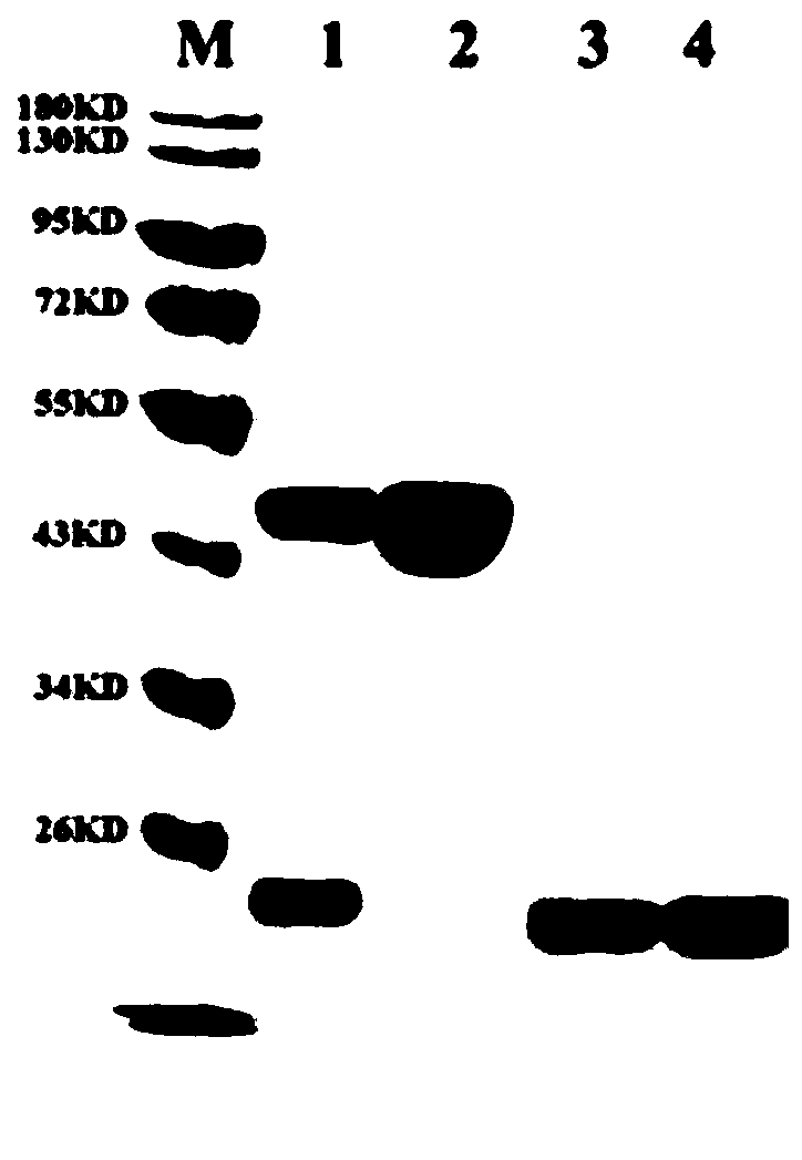
[0044] Embodiment 1, expression and purification of APOBEC3A enzyme

[0045] This application uses pET41a as the vector framework, amplifies the MBP-Xa tag from pMal-c5x, transfers it to pET41a through homologous recombination, and then connects the APOBEC3A gene and the vector to obtain the recombinant plasmid pET41a-MBP-A3A-His. Transform E.coli BL21(DE3)pLysS competent cells (Tiangen Biochemical) with the recombinant plasmid, and pick 10-20 small positive clones into 5mL liquid LB medium (containing 2.5mM glucose, 50ug / mL Kanamycin in chloramphenicol, 34ug / mL chloramphenicol, 100uM EDTA), 37°C, 250rpm to OD600 of about 0.5, quickly cooled on ice, then added IPTG to a final concentration of 0.2mM, and cultured overnight at 16°C. Collect the bacteria by centrifugation, resuspend the bacteria in 0.5ml PBS, add lysozyme and incubate at 37°C for 30min, and take the supernatant for Western Blot analysis. The clones whose expression was confirmed by WB were scaled up and cultured...

Embodiment 2

[0046] Example 2, verification of the deamination activity of APOBEC3A

[0047] 1. DNA substrate preparation: Amplify a DNA fragment from plasmid DNA by nested PCR, 580bp in length;

[0048] 2. APOBEC3A deamination conversion process

[0049] (1) Prepare APOBEC3A Enzyme Mix on ice according to the following system, shake slightly to mix evenly, centrifuge briefly and insert on ice for later use.

[0050] Table 1

[0051]

[0052]

[0053] (2) DNA denaturation: Mix the components according to the following system, insert on ice to cool for 1 min, transfer to a 95°C PCR instrument for denaturation for 3 min, quickly place in an ice-water bath, and let stand for 2 min.

[0054] Table 2

[0055]

[0056] (3) Quickly mix the Enzyme Mix and the denatured product, gently blow and mix with a pipette gun, centrifuge briefly at 4°C and place on ice for later use.

[0057] (4) Transfer the above-mentioned mixed product to a PCR instrument to start the transformation program....

Embodiment 3

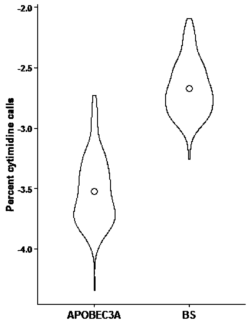
[0067] Example 3, using APOBEC3A to distinguish 5-mC and 5-hmC

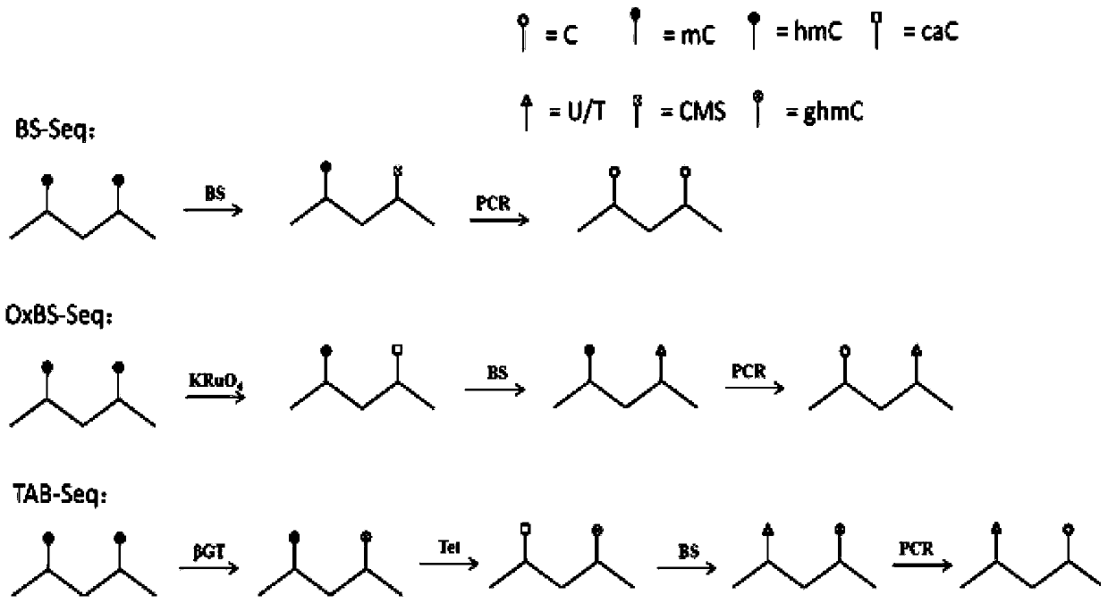
[0068] This example provides a method for quickly distinguishing 5-mC and 5-hmC based on the difference of APOBEC3A in the deamination conversion of 5-mC and 5-hmC. The principle is as follows Figure 4 shown.

[0069] 1. DNA substrate preparation

[0070] Use 5-mC dCTP instead of dCTP for PCR amplification, a DNA fragment amplified from λDNA is named mC-λ3, and a DNA fragment amplified from pMal-c5x_RH2 is named mC-RH2; use 5-hmC dCTP instead of dCTP PCR amplification, a DNA fragment amplified from λDNA was named hmC-λ3, and a DNA fragment amplified from pMal-c5x_RH2 was named hmC-RH2.

[0071] 2. Glycosylation of 5-hmC

[0072] Table 4 reaction system

[0073]

[0074]

[0075] Reaction conditions: add each component in order, mix well, transfer to PCR machine, incubate at 37°C for 1h, hold at 4°C, heat cover at 50°C.

[0076] Product recovery: After the reaction, use Oligo Clean&Concentrator (Zymo R...


PUM


Abstract

The invention belongs to the technical field of epigenetics and particularly relates to a method for differentiating 5-methylcytosine (5-mC) and 5-hydroxymethylcytosine (5-hmC) in DNA. According to the method, based on the difference of APOBEC3A on deamination transformation of 5-mC and 5-hmC, a detected site is directly used as the 3' end of a specific primer, different amplification Ct values can be generated by different methylation modifications after qPCR amplification, and whether the modification of the detected site is methylation modification or hydroxymethylation modification can bequickly differentiated according to the Ct value difference between an enzymatic transformation product and a negative control, so that the analysis timeliness can be improved, and the cost for secondary sequencing can be saved. The method almost has no damage on a DNA in a transforming process, can complete analysis by utilizing a sample DNA as low as 1ng, and can be widely applied to the field of DNA epigenetic analysis.

Description

technical field [0001] The invention belongs to the technical field of epigenetics, and in particular relates to a method for distinguishing 5-methylated cytosine and 5-hydroxymethylated cytosine in DNA. Background technique [0002] The methylation modification at the C5 position of cytosine is one of the most common DNA modification methods in nature, and it is of great significance in the fields of chromosome inactivation, gene regulation, embryonic development, and disease control. mC) is known as the "fifth base" in nature. In recent years, with the continuous research on 5-methylated cytosine, scientists have discovered the "sixth base" in nature: 5-hydroxymethylated cytosine (abbreviated as 5-hmC). In fact, this base was discovered in 1952, but it was not taken seriously until 2009, when two scientists, Kriaucionis and Tahiliani, confirmed the existence of 5hmC in Purkinje cells, granule neurons and embryonic stem cells of mice (Kriaucionis and Heintz, 2009; Tahilia...

Claims


Application Information

Patent Timeline
IPC IPC(8): C12Q1/6806C12Q1/6858
CPCC12Q1/6806C12Q1/6858C12Q2521/539C12Q2521/537C12Q2527/125C12Q2527/101C12Q2531/113C12Q2521/531
Inventor 刘震李阳
Owner WUHAN BIORUN BIO TECH
Why Eureka
Try Eureka