Method for detecting anti-AQP4 antibody based on quantum dot polystyrene microsphere
A technology of polystyrene microspheres and quantum dots, applied in the fields of bioanalytical chemistry and nanobiology, can solve the problems of low protein expression affecting detection results, difficulty in realizing large-scale commercial production, and difficulty in establishing quality control standards, etc., to achieve Rapid detection of standardization and automation, easy standardization and automation, wide excitation spectral range effects
- Summary
- Abstract
- Description
- Claims
- Application Information
AI Technical Summary
Problems solved by technology
Method used
Image
Examples
Embodiment 1
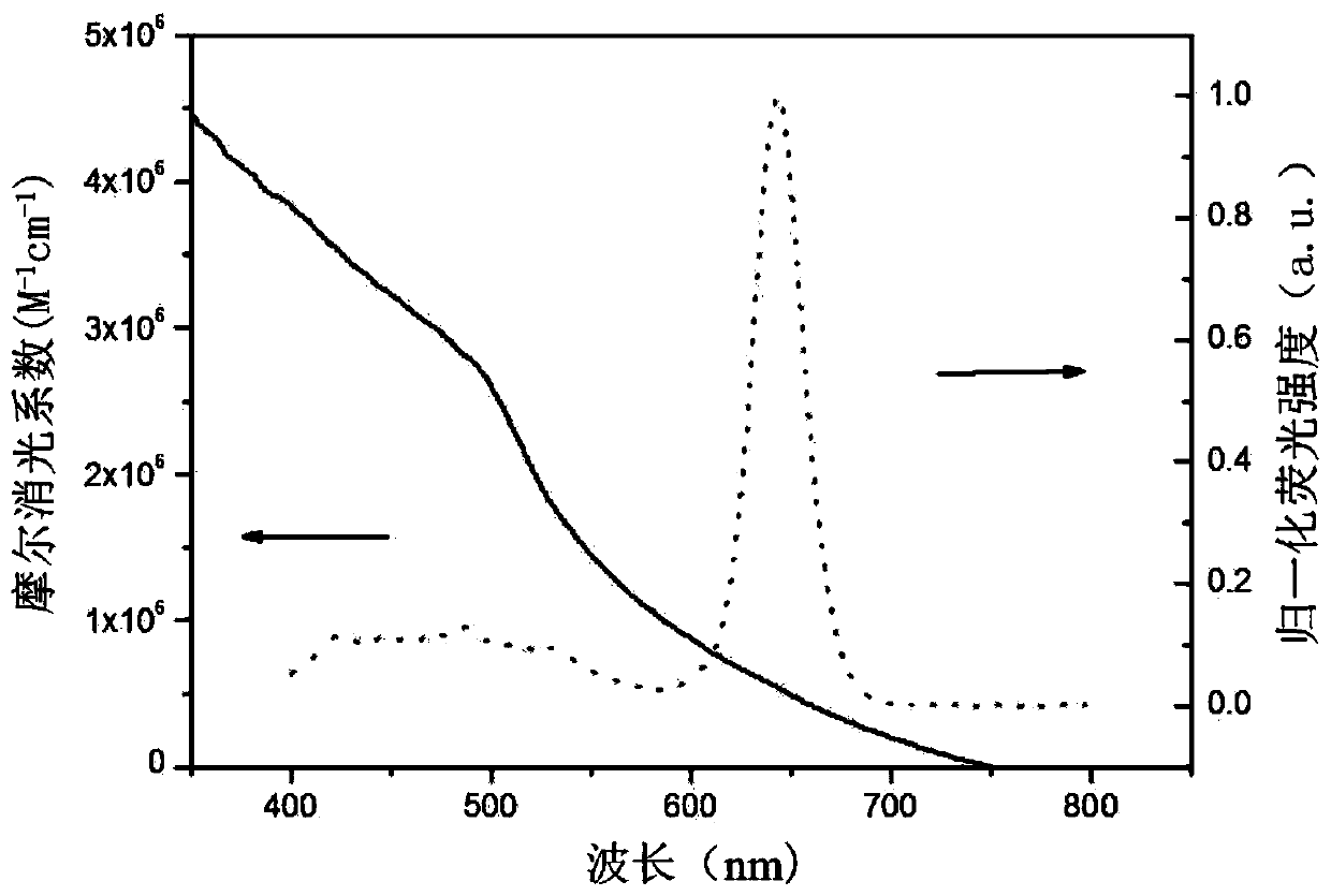
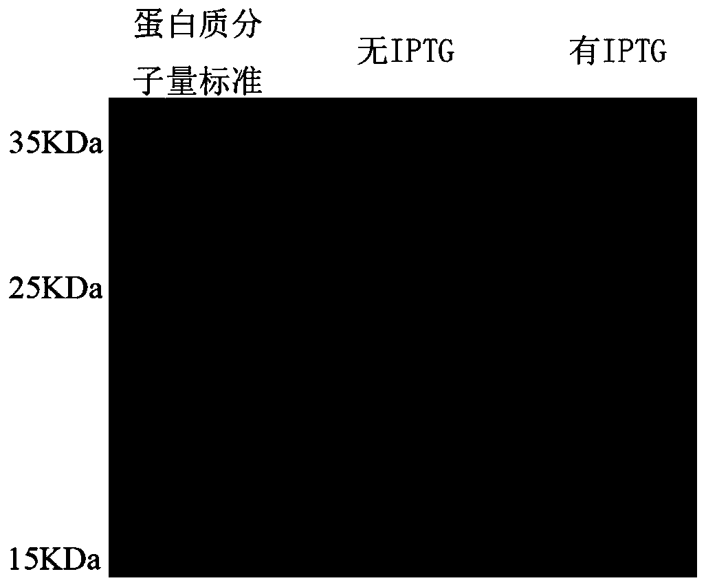
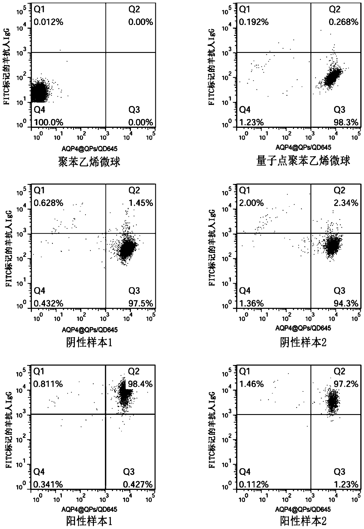
[0054] A method for detecting anti-AQP4 antibodies based on quantum dot polystyrene microspheres, such as Figure 1 to Figure 3 shown, including the following steps:
[0055] (1) Preparation of quantum dot polystyrene QPs microspheres;
[0056] (2) obtaining the functional fragment target gene of AQP4 protein for expression;
[0057] (3) Utilize EDC-NHS to activate the carboxyl groups on the surface of the QPs microspheres, and couple the carboxyl groups on the surface of the activated QPs microspheres with the amino groups of the AQP4 protein to make functionalized QPs microspheres;
[0058] (4) Flow cytometric detection by oil-soluble CdSe quantum dots with a fluorescence emission peak of 600nm to 650nm and FITC-labeled goat anti-human IgG red-green dual fluorescence localization system, and the detection results of the functionalized QPs microspheres to read.
[0059] Specifically, step (1) adopts chemical osmosis method to prepare QPs microspheres, and the specific proc...
Embodiment 2
[0098] A method for detecting anti-AQP4 antibodies based on quantum dot polystyrene microspheres, other features are the same as in Example 1, the difference is that the specific detection steps of the detection sample are as follows:
[0099] (1) Take 10 μL serum sample, dilute it with PBS solution according to the ratio of serum: PBS solution = 1:10, fully mix the diluted serum with 80 μg of QPs-AQP4 complex, and incubate at 37°C for one hour.
[0100] (2) Wash the incubated product 3 times with PBS solution, 5 minutes each time, and spin dry.
[0101] (3) Dilute FITC-labeled goat anti-human IgG with PBS solution according to the ratio of FITC-labeled goat anti-human IgG: PBS solution = 1:50, and add 100 μL of diluted FITC-labeled goat anti-human IgG to every 100 μL serum mixture sample , and incubate the mixture at 37°C for one hour in the dark.
[0102] (4) Wash the incubated functionalized QPs microspheres-anti-AQP4 antibody-goat anti-human IgG immune complex three times...
Embodiment 3
[0107] A method for detecting anti-AQP4 antibodies based on quantum dot polystyrene microspheres, comprising the following steps:
[0108] (1) Preparation of quantum dot polystyrene QPs microspheres;
[0109] (2) obtaining the functional fragment target gene of AQP4 protein for expression;
[0110] (3) Utilize EDC-NHS to activate the carboxyl groups on the surface of the QPs microspheres, and couple the carboxyl groups on the surface of the activated QPs microspheres with the amino groups of the AQP4 protein to make functionalized QPs microspheres;
[0111] (4) The red-green dual fluorescence localization system of oil-soluble CdSe quantum dots with a fluorescence emission peak of 600nm and FITC-labeled goat anti-human IgG was used for flow cytometry detection, and the detection results of the functionalized QPs microspheres were interpreted.
[0112] Specifically, step (1) adopts chemical osmosis method to prepare QPs microspheres, and the specific process includes:
[0113...
PUM
 Abstract
Description
Claims
Application Information
 - R&D
- Intellectual Property
- Life Sciences
- Materials
- Tech Scout
- Unparalleled Data Quality
- Higher Quality Content
- 60% Fewer Hallucinations
Browse by: Latest US Patents, China's latest patents, Technical Efficacy Thesaurus, Application Domain, Technology Topic, Popular Technical Reports.
© 2025 PatSnap. All rights reserved.Legal|Privacy policy|Modern Slavery Act Transparency Statement|Sitemap|About US| Contact US: help@patsnap.com


